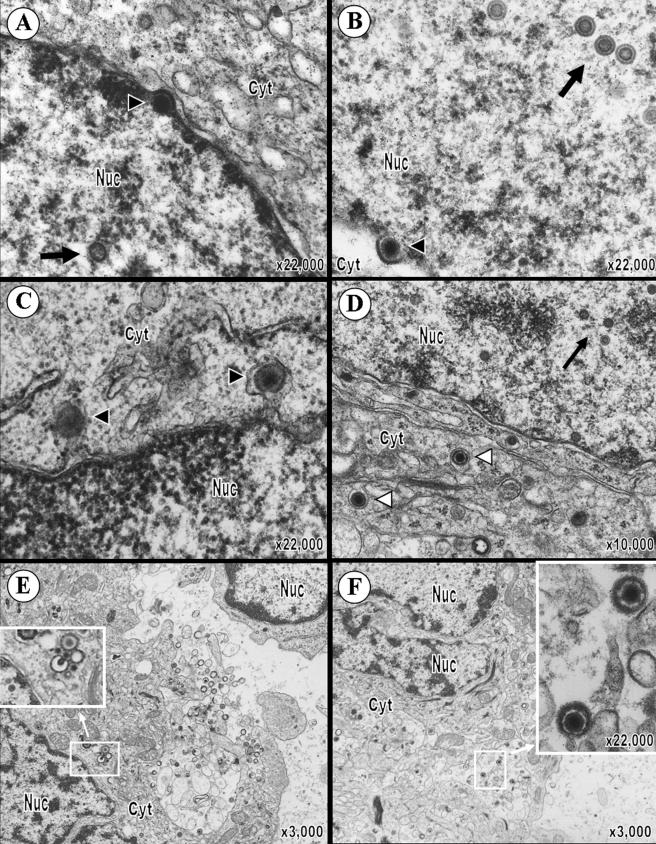
FIG. 5.
Perinuclear and cytoplasmic localization of pOka virions within human T cells in SCID-hu T-cell xenografts. VZV nucleocapsids can be seen budding at the inner nuclear membrane (A and B). Arrowheads point to budding virions. In panel A, the indicated budding nucleocapsid is adjacent to the inner leaflet of the nuclear membrane and the site of budding is marked by an area of increased electron density. In panel B, the budding viral particle is nearly completely surrounded by nuclear membrane. (C) Virions in the perinuclear cleft (indicated by black arrowheads) appear to have a tegument and envelope. (D) Complete enveloped virions can be seen within vesicles in the cytosol (white arrowheads) and are significantly larger than capsids in the nucleus (large arrows). (E) The inset shows the magnification of a nucleocapsid in the cytoplasm budding into a cytoplasmic vesicle. Next to this is a complete virion, an empty vesicle, three capsids containing a DNA core, and an empty capsid. Virions in various stages of maturation can be seen throughout the cell, as well as exiting the plasma membrane from a cytoplasmic vesicle. (F) Complete virions within a large cytoplasmic vesicle consist of a viral DNA core, capsid, tegument, envelope, and protein spikes (inset). The magnification and locations of cytoplasm (cyt) and nucleus (nuc) are indicated.

