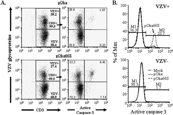
FIG. 7.
Flow cytometric analysis of apoptosis in pOka- and pOka66S-infected T cells. Purified human tonsil T cells were infected with either pOka or pOka66S and stained at 48 h postinfection for VZV proteins, CD3, and active caspase 3. (A) Cells cultured with pOka-infected fibroblasts (top panels) or pOka66S-infected fibroblasts (bottom panels) were gated on CD3-positive T cells (gating shown in left panels). Only 1% of all T cells cocultured with pOka-infected cells expressed active caspase 3 (top right panel), compared with more than 6% of T cells cocultured with pOka66S (bottom right panel). (B) T cells were divided into VZV-positive (top panel) and VZV-negative (bottom panel) populations. Over 20% of the T cells infected with pOka66S had detectable active caspase 3 (top panel), whereas uninfected T cells from the same culture were almost entirely negative for the presence of active caspase 3 (bottom panel), as were those infected with pOka (top panel).

