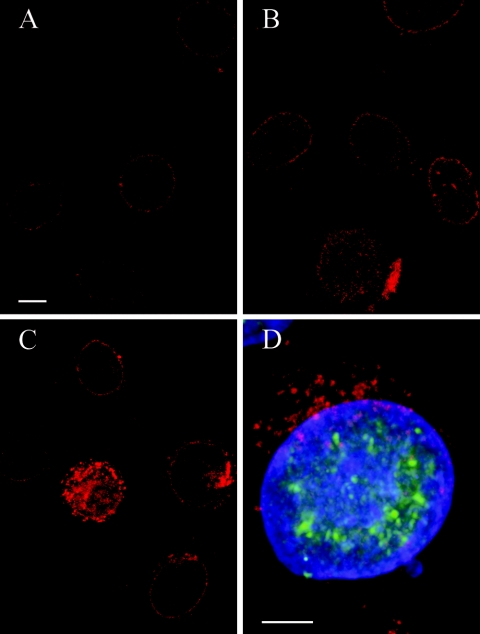
FIG. 4.
Confocal microscopy of HeLa cells after immunolabeling of pore complex protein Nup153 (Texas Red) and of HSV-1 immediate-early protein ICP4 (D) and subsequent DAPI staining. (A) Faint Texas Red signals are regularly distributed at the nuclear surface in cells incubated for 4 h with HSV-1. (B) Signal intensity is increased at the nuclear surface in cells incubated for 6 h. (C) Irregular accumulation of Texas Red signals at the nuclear periphery and in the cytoplasm at 8 h postinfection. (D) Merger of Texas Red, FITC, and DAPI staining demonstrating that dislocation of Nup153 from the nuclear periphery into the cytoplasm occurred only in cells expressing HSV-1 virus protein ICP4 (FITC). Bars, 5 μm.

