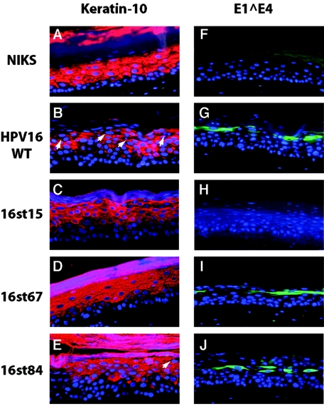
FIG. 5.
Analysis of E1∧E4 expression and cellular differentiation in raft cultures. Cross sections of raft cultures were subjected to immunofluorescence with antibodies to keratin-10 (A to E) (red) or E1∧E4 antibody (F to J) (green) and counterstained with DAPI to indicate nuclei (blue), as indicated in Materials and Methods. Arrows indicate the K10-negative cells in the more superficial spinous and granular layers. Note that monoclonal antibodies used to detect E1∧E4 recognize an epitope not present in the 16st15 E1∧E4 gene product. Shown are panels from rafts of NIKS cells alone (A and F), NIKS cells harboring the wild-type genome (B and G), or NIKS cells harboring the indicated E4 mutant HPV16 genomes (C to E and H to J).

