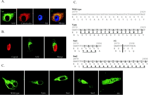
FIG. 3.
The localization of SAT. (A) SAT localizes in the ER. The pFLAG68 viral construct expressing FLAG-labeled SAT was cotransfected with the pDsRed2-ER vector expressing the ER marker calreticulin fused to DsRed2 fluorescent protein. Anti-FLAG (mouse) and anti-NS1 (rabbit) antibodies were labeled with Alexa Fluor-488 and Alexa Fluor-647 giving green and blue fluorescence, respectively. The extensive colocalization of SAT and calreticulin resulted in orange-yellow staining in the merged image. (B) SAT does not colocalize with the viral capsid proteins. The pSATins68 viral construct expressing GFP-labeled SAT was cotransfected with the infectious clone of the wild-type virus pN2D. Anti-GFP and anti-capsid antibodies were labeled with Alexa Fluor-488 and Alexa Fluor-568. (C) Changes in the G-stretch sequence of mutants derived from pSATIns68. The changes in the nucleotide and protein sequences are boldface type and underlined; the nucleotide sequences are numbered according to their original position in the PPV NADL-2 strain. The position of deletion in the ΔG clone is indicated by a vertical bar. (D) Localization of the G-stretch-modified SAT proteins. The G-stretch mutant constructs listed in panel C were transfected and the expression of SAT-GFP fusion proteins was followed with Alexa Fluor-488-labeled anti-GFP antibody. Images are labeled with the names of the corresponding mutants.

