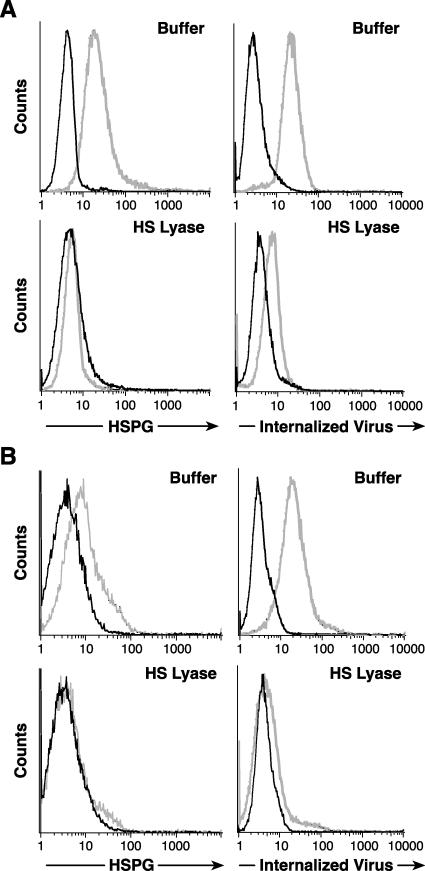
FIG. 7.
HSPGs enhance HTLV-1 Env-mediated entry into CD4+ T cells. (A) MOLT4 cells were incubated either with (bottom) or without (top) 20 mU of HS lyase as described in the legends to the previous figures. (Left) HSPG expression was determined as described above. Dark lines, isotype control; light lines, F58-10E4. (Right) The extent of internalization was determined 2 h after exposing the cells to HTLV-1 virions, as described in Materials and Methods. Dark lines, mouse IgG1 (isotype control); light lines, anti-HTLV MA (p19) antibody. Without HS lyase treatment, the MFI was 17.8, and 89% of the cells were positive for internalized virus. With HS lyase treatment, the MFI was 2.6, and 8.9% of the cells were positive. (B) CD4+ T cells were isolated from cord blood lymphocytes, activated for 3 days with anti-CD3/anti-CD28 antibody beads, and then treated with 10 mU of HS lyase (bottom) or left untreated (top). (Left) HSPG binding to F58-10E4 (light lines) or isotype control (dark lines). (Right) Binding of anti-MA (p19) antibody (light lines) or isotype control (dark lines). The data shown are from a representative experiment out of 14 performed.

