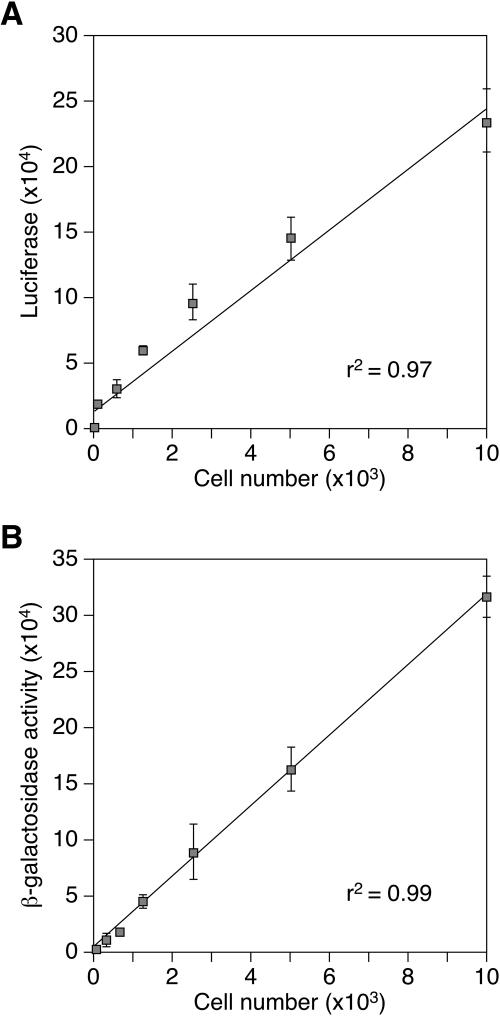
FIG. 2.
The β-galactosidase-based infection assay and the luciferase-based cell viability assay display a linear dependence upon cell number. Cells were plated for 24 h and then challenged with 8 × 10⁴ LTU of MMP-nls-lacZ[VSV-G]. The cells were assayed 48 h postinfection with (A) a CellTiter-Glo kit (Promega, Madison, WI), which measures the viability of infected cells by measuring cellular ATP as a substrate for luciferase, or (B) a Galacto-star chemiluminescent β-galactosidase assay (Applied Biosystems, Foster City, CA). Uninfected controls were assayed 72 h after plating. The data shown are the average mean values obtained in an experiment with quadruplicate samples and are representative of results of three independent experiments. Error bars indicate the standard deviations of the data.

