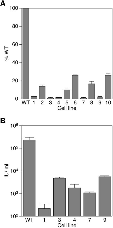
FIG. 3.
Resistance of isolated cell lines to infection by a VSV-G-pseudotyped MLV vector. (A) Cells (1 × 10⁴/well of a 96-well plate) of the indicated cell lines were infected with 5 × 10⁴ IU of MMP-nls-lacZ[VSV-G] and assayed 48 h postinfection with chemiluminescent assays for β-galactosidase and viability as described for Fig. 2. (B) Cells (1 × 10⁴/well) of the indicated cell lines were infected with different concentrations of MMP-nls-lacZ[VSV-G] and stained 48 h postinfection with X-Gal. The data shown are the average mean values obtained in an experiment with triplicate samples and are representative of results of three independent experiments. Error bars indicate the standard deviations of the data.

