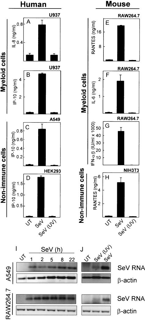
FIG. 1.
Induction of cytokine expression by SeV. U937 (A and B), A549 (C), HEK293 (D), RAW 264.7 (E to G), and NIH 3T3 (H) cells were seeded in 96-well plates and left overnight to settle. The cells were left untreated (UT) or treated with infectious (SeV) or UV-inactivated [SeV (UV)] SeV at a multiplicity of infection (MOI) of 1. Supernatants were harvested 16 to 24 h later, and cytokine levels were measured by ELISA or bioassay. The results are shown as means of triplicate cultures ± SEM. Essentially similar results were observed for two to five independent experiments. (I and J) A549 and RAW 264.7 cells were seeded and left overnight to settle prior to treatment with infectious and UV-inactivated SeV at an MOI of 1. After the indicated times of infection, total RNA was harvested and accumulation of viral RNA and cellular β-actin was assessed by RT-PCR. Similar results were obtained for two independent experiments.

