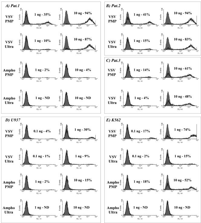
FIG. 3.
Comparative infectivity of lentivirus in primary and established leukemia cells after concentration by either PMP or ultracentrifugation. Lentiviral vectors expressing enhanced green fluorescent protein (LV.gfp) were prepared from stable B7.1-expressing 293T cells (VSV-G) or 293T-Ampho (Ampho) and concentrated 100-fold either by CTLA4-Ig-conjugated PMP capture (PMP) or by ultracentrifugation (Ultra). After enzyme-linked immunosorbent assay determination of p24 Gag the viral concentrates were used at equimolar p24 levels to infect three cryopreserved primary AML samples (A to C; patients 1 to 3, respectively) and the established leukemia cell lines U937 and K562 (D and E, respectively) all in the presence of 4 μg/ml Polybrene. Fluorescence-activated cell sorting analysis of enhanced green fluorescent protein expression was carried out 96 h after infection (black line, enhanced green fluorescent protein; shaded area, background). No enhanced green fluorescent protein expression was detected for primary AML samples 2 and 3 following inoculation with amphotropic virus concentrated by either strategy (data not shown). ND, not detected.

