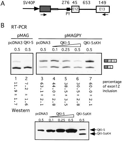
Figure 2.
The repression of MAG exon 12 inclusion by QKI-5. (A) Schematic drawing of the MAG minigene construct. A 1.1-kb genomic DNA fragment around MAG exon 12 is cloned downstream of an HIV tat exon (gray box) in the vector pSPL3. SV40P, simian virus 40 promoter. The numbers above refer to the size (bp) of corresponding exon or intron. The arrows represent the primers used in the RT-PCR in B. The polypyrimidine (PY) tract of exon 12 is indicated by the small black box. In the pMAGPY construct, it was replaced by the PY of exon 11 (from CGTTTCCTTCTTCAAT to ATGTTTCATGCCTCCCTGC). (B) Representative RT-PCR results of RNA from COS7 cells cotransfected with 0.5 μg of MAG minigene and indicated amounts of QKI expression plasmid. The vector pcDNA3 was used to maintain the total level of transfected DNA and as a negative control. The intensity of the bands was measured with a PhosphorImager, and the percentage of exon 12 inclusion is marked below each lane with standard deviations (n = 3). A Western blot with anti-QKI-5 antibody indicates the expression of QKI-5 and QKI-5ΔKH proteins in cells cotransfected with pMAGPY and indicated amounts of QKI-5 expression constructs. The endogenous QKI-5 protein was indicated by the band in the pcDNA3 only lane.

