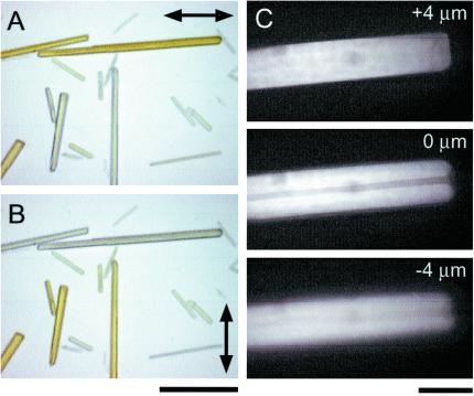
Figure 3.
(A and B) Dichroism of GFP crystals observed with quartz halogen illuminator. Double-headed arrows: polarizer transmission axis. (Bar = 30 μm.) (C) Confocal epifluorescence optical sections taken 4 μm above, at, and 4 μm below the mid plane of GFP crystal with large hollow core. The asymmetry of the image above and below the midplane and the strong flare reflects the high refractive index of the crystal wall material. (Bar = 10 μm.)

