Figure 7.
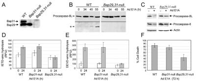
Procaspase-8L processing is inhibited in Bap29,31-null cells. (A) Loss of Bap29 and Bap31 expression was confirmed by immunoblotting with anti-BAP31 and anti-Bap29 antibodies. (B) E1A-induced procaspase-8L cleavage is inhibited in Bap29,31-null ES cells. ES cells were differentiated into epithelial- and fibroblast-like cells as described in Materials and Methods, and infected with Ad5 dl520E1B− for the indicated times. Procaspase-8L cleavage was analyzed as in Fig. 3A. (C) As in B, procaspase-8L and procaspase-8/a and -8/b cleavage was analyzed with anti-Nex and anti-caspase-8 (p18) antibodies. (D) Decreased caspase-8 activity in Bap29,31-null cells. Aliquots of lysate from wild-type and Bap29,31-null cells (containing equivalent protein concentrations) stimulated with E1A for 24h (time of maximum caspase activity) were tested for their ability to hydrolyze the preferred caspase-8 substrate IETD-amc. Shown is the average of three independent experiments. RFU, relative fluorescence units. (E) Decreased DEVD-ase activity in Bap29,31-null cells. As in C, except that lysates were tested for their ability to hydrolyze the caspase-3 preferred substrate DEVD-amc. (F) Cell death was measured by trypan blue exclusion 72 h post infection. Shown is a representative of four independent experiments.

