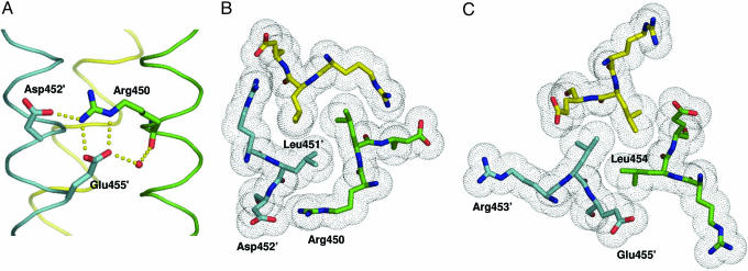
Fig. 2.
Prominent structural motif seen in the ccCor1 trimer. (A) Side view of the salt-bridge network (indicated by yellow dots) formed between Arg-450, Asp-452′, and Glu-455′ and the water-mediated hydrogen bond between Glu-455′:Oε2 and Arg-450:O. (B) End-on view of the a3 layer showing the shielding of the Leu-451 residues from solvent by the aliphatic side-chain moieties of Arg-450. (C) End-on view of the d3 layer showing the hydrophobic packing between the Leu-454 and the aliphatic side-chain moieties of the Glu-455 residues. Side chains of residues are shown as stick representation and van der Waals spheres (B and C), the water molecule as a small red sphere (A), and the three monomers are shown as Cα traces. Colors of atoms and monomers are the same as in Fig. 1.

