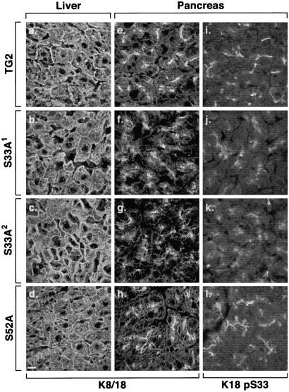
Figure 2.
Immunofluorescence staining of keratins in transgenic mouse tissues. Liver and pancreas were isolated from the indicated transgenic lines. Tissues were freshly frozen, sectioned, and then fixed in cold acetone. Staining was performed with mAb L2A1, which recognizes the hK18 transgene product (a–h) and anti-K18 pS33 (i–l). Note that the “tight” apicolateral filament distribution, particularly noted in i and l (pancreata of TG2 and S52A mice, respectively), are more dispersed in S33A mice pancreata (j and k; similar findings were noted with anti-hK18 pS52 Ab, not shown). (Bar = 100 μm.)

