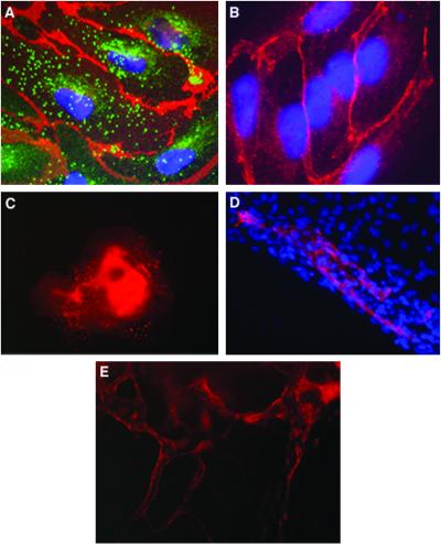
Figure 2.
Expression of endothelial cell markers in vessel-like structure within hEBs. (A) EBs at day 13 stained with human PECAM1 antibodies (red), vWF antibodies (green), and DAPI for nuclear staining (blue). PECAM1 is organized at cell–cell junctions, whereas vWF is found in organelles in the cytoplasm. (B) EB cells stained with human VE-cadherin antibodies (red) and DAPI (blue) (magnification, ×1,000). (C) Low magnification (×100) of EB stained with PECAM1 antibodies. (D) Areas of PECAM1+ cells (red) within part of an EB, organized in elongated clusters. Cell nuclei stained with DAPI (blue) (magnification, ×400). (E) Channels forming PECAM1+ cells within a 13-day-old EB (magnification, ×200).

