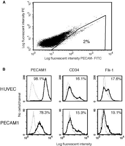
Figure 4.
Isolation of endothelial cells from human embryoid bodies by using fluorescent-labeled anti-PECAM1 antibodies and analysis of the sorted cells. (A) EBs at day 13 were dissociated and incubated with PECAM1 antibodies. Fluorescent-labeled cells were isolated by using a flow cytometry cell sorter. (B) Flow cytometric analysis of endothelial cell markers in PECAM1+ cells grown in culture for six passages and HUVEC cells. The cells were dissociated and incubated with either isotype control (dashed lines) or antigen-specific antibodies, as indicated (solid lines). Percent positive cells are shown.

