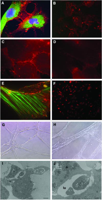
Figure 5.
Characterization of hES-derived endothelial cells grown in culture. (A) Immunofluorescence staining of PECAM1 (red) at cell–cell junctions and vWF (green) in the cytoplasm. The nuclei are stained with DAPI (blue). (B) Cells stained for PECAM1. (C) N-cadherin and (D) VE-cadherin staining in cell–cell adherent junctions. (E) Double staining for vinculin (red) and actin (green). Vinculin is found in both focal contacts and cell–cell adherent junctions where it associates with actin stress fiber ends (magnification: A and C–E, ×1,000; B, ×200). (F) Uptake of Dill-labeled ac-LDL by PECAM1+ cells. (G and H) Cords formation by PECAM1+ cells 24 h (G) or 3 days (H) after seeding the cells in matrigel (magnification: G, ×100; H, ×200). (I) Electron microscopy of the cord cross-section showing lumen formation (bar = 2 μm) and (J) higher magnification of the lumen (lu) area showing cell–cell interactions closing the lumen and the nucleus (n) of one cell (bar = 8 μm).

