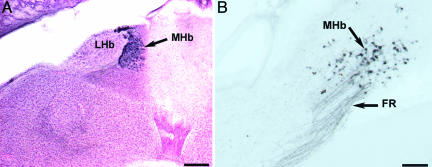
Fig. 4.
The MHb in coronal sections. (A) Neutral red and ChAT-IR (dark blue) cells show the location of the MHb between the lateral habenula on the left (lateral) and the anterior-most portion of the cerebellum on the right. Dorsal is up. (Scale bar, 200 μm.) (B) Retrograde-labeled cells from tracer injections into the ipsilateral Uva show labeled axons in the fasciculus retroflexus and cells in the MHb. Dorsal is up, and medial is to the right. (Scale bar, 50 μm.)

