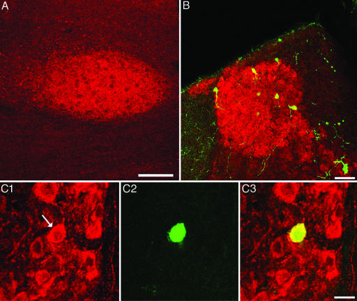
Fig. 6.
The MHb provides cholinergic input to the Uva. (A) Coronal section of an adult male zebra finch shows ChaT-IR in the Uva. Dorsal is up, and lateral is to the left. (Scale bar, 200 μm.) (B) Low-power view of the MHb double labeled with ChAT-IR (red) and retrograde cells from an ipsilateral Uva injection of a fluorescent green tracer. Dorsal is up, and medial is to the right. (Scale bar, 50 μm.) (C) High magnification of cells double labeled in the MHb. (C1) ChaT-IR cells in the MHb; arrow points to a cell of interest. (C2) The same cell is retrogradely labeled from the Uva with a green tracer. (C3) An overlay of C1 and C2 confirms that this cell is double labeled. Dorsal is up, and medial is to the right. (Scale bar, 20 μm.)

