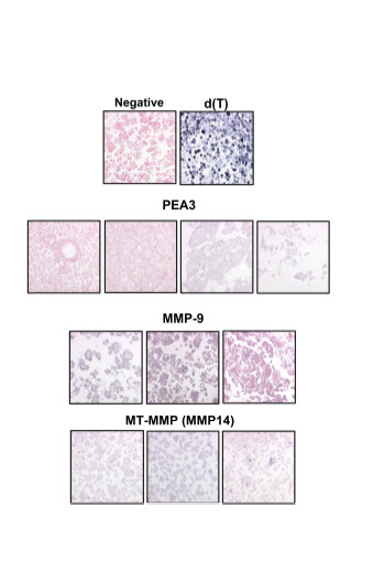
Figure 3.

In situ hybridization of selected genes. mRNA in situ hybridization in ovarian carcinoma effusions. Negative control specimen (stained with nuclear fast red) and d(T) control are shown in the first row. Two negative (left, stained red) and two positive (right, gray) cases using the PEA3 probe are shown in the second row. Two positive (left, stained gray) and one negative (right, red) cases using the MMP-9 probe are shown in the third row. Three positive cases using the MMP14 probe are shown in the fourth row.
