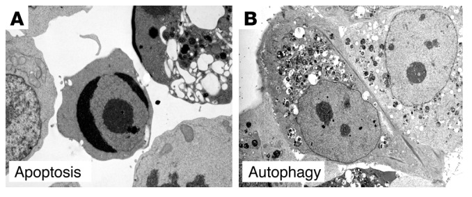
Figure 3.

Ultrastructural examples of apoptotic and autophagic cell death. Electron micrographs of a FasL-treated Jurkat cell undergoing cell death with apoptotic features (A) and of a tamoxifen-treated MCF7 human breast carcinoma cell undergoing cell death with autophagic features (B). In A, note chromatin condensation (cell in center) and cytoplasmic vacuolization (cell in upper right). In B, note absence of chromatin condensation and presence of numerous autophagosomes. Images in A and B reproduced with permission from Nature Cell Biology (98) and Landes Bioscience (90), respectively.
