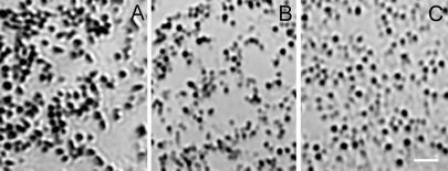
Figure 6.
The cht/cht melanosomes are similar in morphology to Tyrp1b melanosomes. Shown are bright-field images of melanosomes from the periphery of primary cultured melanocytes, isolated from (A) C57BL/6J+/+, (B) C57BL/6J Rab38cht/Rab38cht mice, and (C) Tyrp1b/Tyrp1b melan-b cells. Melanosomes from wild-type melanocytes are oval and darkly pigmented. In contrast, Rab38cht/Rab38cht melanosomes are smaller, more circular and less pigmented, similar to melanosomes from (C) Tyrp1b/Tyrp1b melan-b cells. (Scale bar = 2 μM.)

