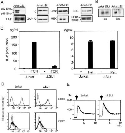
Figure 1.
J.SL1 cells lack expression of Shc and are defective in IL-2 production. (A) Cell lysates of Jurkat and J.SL1 cells were analyzed by Western blots with the antibodies shown on the side of each panel. (B) Northern blot for lck and shc mRNA. Total RNA from Jurkat and J.SL1 cells was analyzed by using the cDNA fragment for lck (Left) or shc (Right). (C) IL-2 production by Jurkat and J.SL1 cells. Cells were cultured 16 h with (TCR) or without (−) stimulation by anti-TCR (Left) or with PMA and ionomycin (P+I, Right). The amount of IL-2 in the culture supernatant from each sample was determined by ELISA. A representative result of several experiments is shown. (D) Surface expression of CD69 and CD25 on Jurkat and J.SL1 cells. Jurkat and J.SL1 cells were treated as described for C, and expression of CD69 and CD25 were examined by using FITC-conjugated antibodies. The dark lines show fluorescence levels detected from activated cells, and gray lines show that from unstimulated cells. (E) Induction of intracellular Ca2+ in Jurkat and J.SL1 cells. Jurkat (Left) and J.SL1 (Right) cells were loaded with Fura-2 acetoxymethyl ester, and the cells were stimulated with anti-TCR at the times shown by the arrow above each panel. The level of Ca2+ was determined on the basis of the ratio between fluorescence from excitation at 340 nm to that from excitation at 380 nm.

