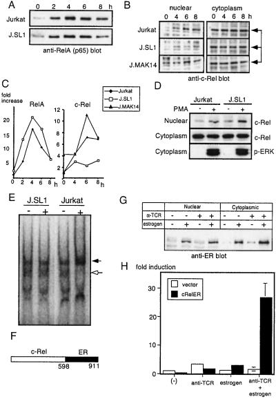
Figure 5.
Reconstitution of IL-2 promoter activity by J.SL1 cells with activation of the c-Rel–ER fusion protein. (A) Nuclear localization of RelA in Jurkat and J.SL1 cells. The time course of nuclear localization of RelA was examined for extracts from Jurkat and J.SL1 cells by Western blot. (B) Nuclear localization of c-Rel is reduced in J.SL1 cells but restored in J.MAK14 cells. The nuclear extract (Left) and cytoplasmic protein (Right) were isolated 4, 6, and 8 h after TCR stimulation. Samples were analyzed by Western blotting using anti-c-Rel antibody. (C) Relative amount of RelA and c-Rel in the nucleus. Relative intensities of bands from the data shown in A and B were determined by using NIH image software. The amount of RelA or c-Rel in the nucleus of Jurkat cells at unstimulated stage (0 h) was taken as 1. (D) Nuclear localization of c-Rel induced by PMA. Jurkat and J.SL1 cells were stimulated with PMA for 30 min. Nuclear and cytoplasmic fractions were analyzed by Western blotting using anti-c-Rel antibody (Top and Middle). An equivalent level of ERK activation was confirmed with anti-phospho-ERK blot (Bottom). (E) Gel supershift assay for c-Rel. Nuclear extracts from stimulated (+) and unstimulated (−) Jurkat and J.SL1 cells were mixed with the 32P-labeled NF-IL-2B site containing oligonucleotide and anti-c-Rel antibody. The shifted band (shown by the darkened arrow) was observed only in activated Jurkat and not in J.SL1 cell nuclear extracts. Note the loss of unshifted band in the activated Jurkat extracts (open arrow). (F) A schematic presentation of the c-Rel–ER fusion protein used in this study with amino acid numbers indicated. (G) Nuclear localization of c-Rel–ER. J.SL1 cells were transfected with the expression construct for c-Rel–ER. Twenty-four hours after transfection, cells were treated with anti-TCR antibody or estrogen as indicated by + above each lane. Nuclear extracts and cytoplasmic fractions of transfectants were analyzed by Western blot using anti-ER antibody. (H) Transient IL-2 promoter assay for J.SL1 transfectants with c-Rel–ER. J.SL1 cells were transfected with the expression vector or the c-Rel–ER expression construct along with the IL-2–luciferase reporter construct as indicated. Four hours after transfection, cells were stimulated with anti-TCR antibody alone, estrogen alone, or anti-TCR antibody and estrogen. Luciferase activity was assayed 18 h after stimulation.

