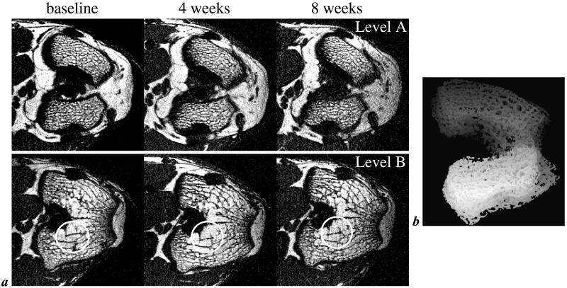
Figure 1.
Typical μ-MR images of trabecular bone. (a) One of 28 contiguous transverse MR images perpendicular to the femoral shaft in the distal femur of the same rabbit at three different time points (image voxel size 98 × 98 × 300 μm3). Locations have been matched to allow comparison of the images collected at baseline and 4 and 8 weeks after s.c. implantation of dexamethasone pellets. Circles highlight structural features well reproduced in the repeat scans. Levels A and B represent locations ≈1 mm distal and 1.5 mm proximal to the intracondylar fossa. Note dense trabecular network in the distal epiphysis (Level A) but sparser trabeculation at the more proximal location (Level B). (b) Representative projection image of the distal femoral epiphysis of a live rabbit covering the volume analyzed. Projection direction is inferior to superior at an angle of 20° relative to the femoral anatomic axis.

