
Figure 2.
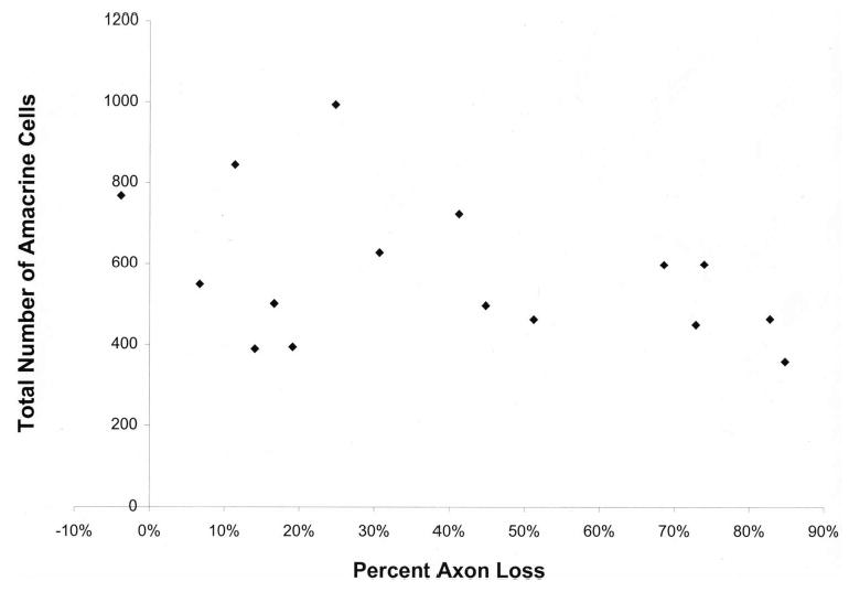
Total amacrine cells labeled by both GABA and glycine in the GCL and INL compared with axon loss in glaucomatous eyes. Amacrine cell count compared with the degree of RGC axon loss in rats with glaucoma shows a statistically non-significant trend toward fewer amacrine cells with greater damage (P = 0.11).