
Figure 4.
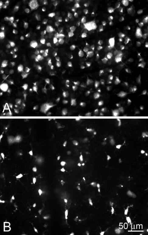
RGC loss after nerve transection. (A) Normal appearance of rat retina with many RGCs that are specifically identified by their morphology, backfilled by injection of fluorescent tracer into the superior colliculus. (B) Two weeks after optic nerve transection, there were no RGCs remaining, and the fluorescent tracer was present within phagocytes that had engulfed the dead RGCs and had a different morphology. Published, with permission, from Blair M, Pease ME, Hammond J, et al. Effect of glatiramer acetate on primary and secondary degeneration of retinal ganglion cells in the rat. Invest Ophthalmol Vis Sci. 2005;46:884–890. © Cadmus Professional Communications.