
Fig. 5.
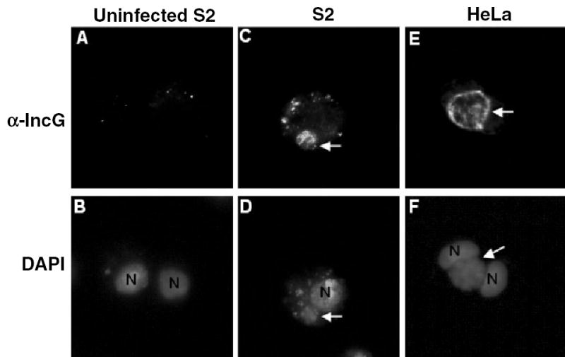
IncG, an RB-specific protein, is expressed in the inclusions of L2-infected S2 cells. S2 and HeLa cells were infected with L2 for 1 h, and then fixed and stained with an IncG monoclonal antibody (upper panels) and DAPI (lower panels) at 48 (S2) or 24 h (HeLa at 37°C) post infection to visualize the inclusion and the host nuclei.
A and B. Uninfected S2 cells.
C–D. S2 cells at 48 hpi.
E and F. HeLa cells at 24 hpi.
Arrows point to inclusions that express IncG, demonstrating that EB to RB differentiation has occurred. N, host nucleus. All micrographs are 1000× final magnification. Samples shown are representatives of multiple fields examined.