


Figure 3.
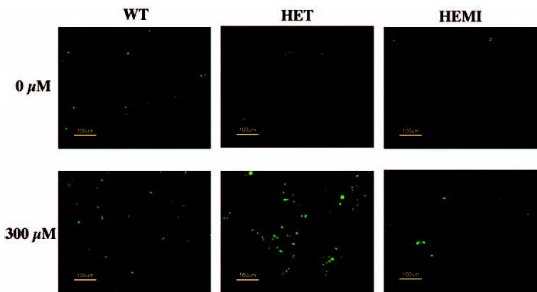
The effect of oxidative stress on apoptosis in RPE cells derived from mice with different levels of SOD2. Cell cultures were exposed to 300 μM H2O2 for 1 hour and recultured for 16 hours in a normal medium, and apoptosis was determined by TUNEL staining. H2O2-treated cells from HET mice showed more TUNEL-positive cells than did those from the WT or HEMI mice, whereas apoptotic cell death was suppressed in the cells from the HEMI mice.