


Figure 5.
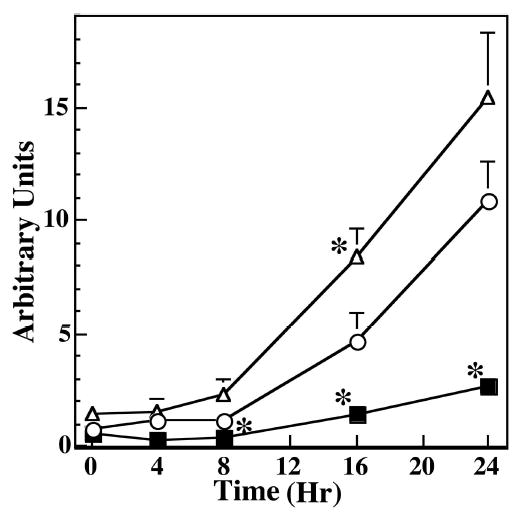
Time course of the effect of H2O2 exposure on apoptosis in RPE with different levels of SOD2. An equal number of RPE cells derived from WT (○), HET (▵), and HEMI (▪) mice were exposed to 300 μM H2O2 for 1 hour and cultured in normal medium for the different periods indicated. Apoptosis was determined by a cell-death–detection ELISA kit. Apoptotic cell death increased progressively as a function of time. Also, greater apoptosis was noted in HET cells than in WT and HEMI cells. Apoptotic cell death in HEMI mice was well suppressed until 16 hours of incubation. Data are expressed as the mean ± SEM (n = 4–7). *P < 0.05 compared with those of WT.