
Fig. 3.
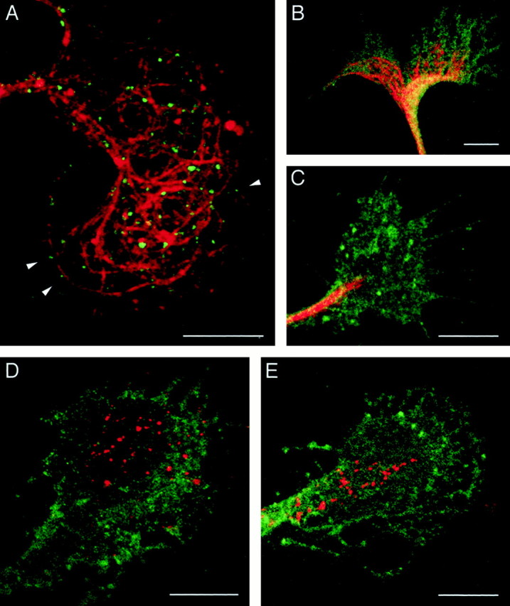
A, Localization of microtubules and endocytosed L1 in a growth cone. DRG neurons cultured on L1 were incubated with anti-L1 antibody for 30 min to allow for internalization of the antibody bound to L1. The cells were fixed and double-labeled for microtubules using an antibody against tyrosinated α-tubulin. Shown is a superimposed image in which endocytosed L1 is colored ingreen and microtubules are colored inred. Arrowheads indicate examples of endocytosed L1 in vesicles positioned along the microtubules. B, C, Effects of taxol on microtubule organization in growth cones. DRG neurons cultured on L1 were pretreated with DMSO (B) or 10 nm taxol (C) for 1 hr and labeled for microtubules using an antibody against tyrosinated α-tubulin (red). The cells were double-labeled for NCAM to outline the growth cone structure (green). D, E, An effect of taxol on the subcellular distribution of endocytosed L1 in growth cones migrating on L1. After pretreatment with DMSO (D) or 10 nm taxol (E) for 1 hr, DRG neurons were incubated with anti-L1 antibody for 30 min to allow for internalization of the antibody bound to L1. The cells were double-labeled for endocytosed L1 (red) and NCAM to outline the growth cone structure (green). Scale bars, 10 μm.