
Fig. 5.
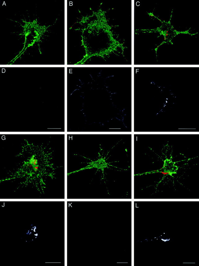
Cell-surface distribution of recycled L1 on growth cones. DRG neurons cultured on L1 (A–G, J) or laminin (H, I, K, L) were allowed to internalize anti-L1 Fab bound to L1 for 30 min, and the cell-surface Fab was blocked. The cells were reincubated for 0 min (A, D, H, K), 30 min (B, E, I, L), 45 min (C, F), or 60 min (G, J) to allow for exocytosis of the L1–Fab complex. Then, recycled L1 was detected by labeling the unblocked Fab that had reappeared on the cell surface. The cells were double-labeled for NCAM to outline the growth cone structure. In superimposed images (A–C, G–I), recycled L1 is colored in red, and NCAM is colored ingreen. To facilitate visualization of recycled L1, thered channel only is shown in black andwhite (D–F, J–L) below the corresponding superimposed image. Scale bars: A, D, 10 μm; B, E, 10 μm; C, F, 10 μm;G, J, 10 μm; H, K, 10 μm; I, L, 10 μm.