
FIG. 5.
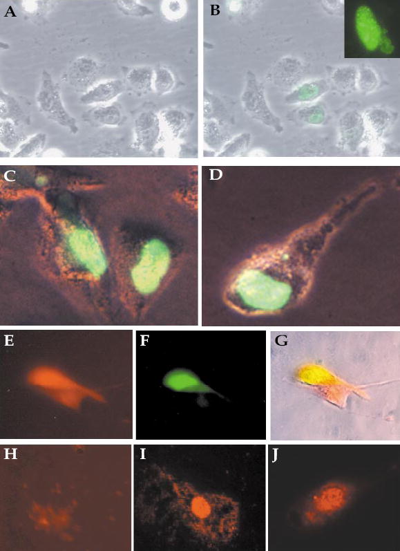
Nuclear localization of mouse INSM1 by immunofluores-cence staining. Untransfected Calu-6 cells (A), Calu-6 cells (B), and HeLa cells (C, D) were transfected with the GFP–INSM1 constructs. The dot-like distribution of GFP–INSM1 in the nucleus was observed by confocal microscopy (inset). (E–G) Calu-6 cells cotransfected with plasmids expressing RFP–CAP (E) and GFP–INSM1 (F). (G) Merge of the two fields. Endogenous INSM1 (I) and CAP (J) proteins detected in the nucleus of HIT-T15 cells stained with antibodies to INSM1 or CAP, respectively. (H) HIT-T15 cells incubated with preimmune serum.