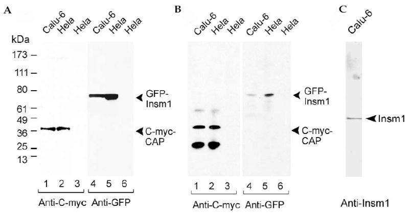
FIG. 7.

Co-immunoprecipitation of mouse INSM1 and CAP in cultured cells. (A) Expression of CAP tagged with c-myc (lanes 1 and 2) and mouse INSM1 tagged with GFP (lanes 4 and 5) in Calu-6 (lanes 1 and 4) and HeLa (lanes 2 and 5) cells. Lanes 3 and 6 represent untransfected cells. Cell extracts were separated on SDS-PAGE, transferred to a nitrocellulose membrane, and stained with antibody to c-myc or GFP. The 37-kDa and 78-kDa bands, respectively, show that CAP and INSM1 were strongly expressed in Calu-6 and HeLa cells. (B) Calu-6 and HeLa cells were co-transfected with c-myc–CAP and INSM1–GFP. Cell lysates were immunoprecipitated with antibody to c-myc coupled to Sepharose beads (or Sepharose beads alone), transferred to a nitrocellulose membrane, and stained with antibody to c-myc or GFP. Lanes 1 and 2 show that immunoprecipitation with antibody to c-myc pulled down c-myc–CAP and lanes 4 and 5 show that GFP–INSM1 was co-precipitated with c-myc–CAP. Lanes 3 and 6 represent untransfected cells. The 61- and 27-kDa bands in lanes 1 and 2 are the heavy and light chains of the antibody to c-myc used for immunoprecipitation. (C) Calu-6 cells that express endogenous INSM1 were transfected with c-myc–CAP. Cell lysates were immunoprecipitated with antibody to c-myc that had been coupled to Sepharose beads, electrophoresed on SDS-PAGE, transferred to a nitrocel-lulose membrane, and stained with antibody to INSM1. Endogenous INSM1 co-precipitated in the c-myc–CAP pull-down.
