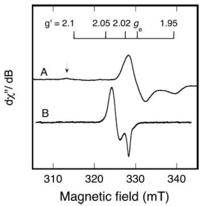
Fig. 5. An EPR spectrum of oxidized RumA compared with the dinitrosyl RumA derivative.

A, the oxidized RumA spectrum (40 K) is the same as that shown in Fig. 3C, but here a low-field region with possible additional absorption is also shown. The arrow indicates minor absorption at ~g′ value = 2.13. B, nitric oxide (from 1 mm nitric oxide donor) was added to deoxygenated RumA (0.17 mm) and the EPR spectrum was recorded (15 K). The fraction of the FeS cluster in the iron-dinitrosyl state, based on double integration, was 33%. Instrument parameters for A are given in the legend to Fig. 3C. For B, the parameters were: microwave frequency, 9.26 GHz; microwave power, 0.1 mW; modulation amplitude, 0.5 mT; sweep rate, 1.3 mT/min; time constant, 0.5 s.
