Figure 2.
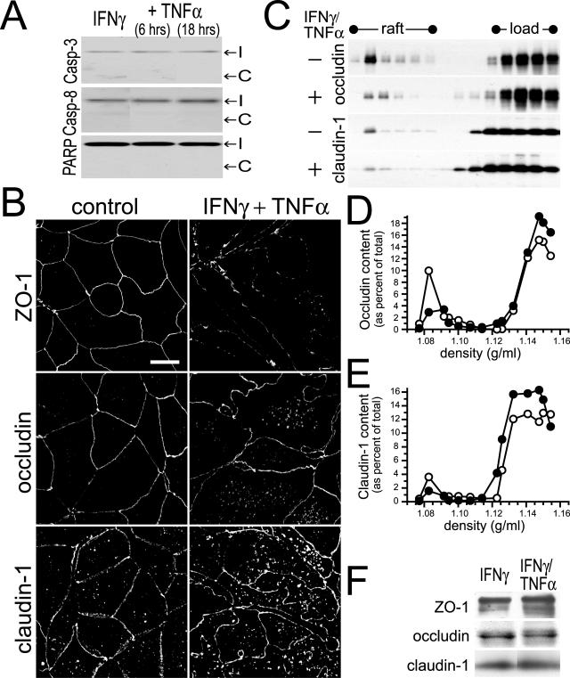
IFN-γ- and TNF-α-induced barrier dysfunction is associated with apoptosis-independent tight junction disruption. A: Caco-2 monolayers were treated with IFN-γ (10 ng/ml) for 24 hours followed by TNF-α (2.5 ng/ml) for times indicated. Cell lysates were analyzed for cleavage of caspase-3, caspase-8, and PARP as markers of apoptosis. No increases in cleaved caspase-3, caspase-8, or PARP were detected in Caco-2 cells treated with the indicated cytokine(s). The positions of intact (I) and cleaved (C) proteins are shown. Data are representative of four similar experiments, each performed in triplicate. B: Caco-2 monolayers were treated with IFN-γ (10 ng/ml) for 24 hours followed by TNF-α (2.5 ng/ml) for 8 hours. Tight junction proteins (ZO-1, occludin, and claudin-1) were detected by immunofluorescence microscopy. Treatment of IFN-γ-primed monolayers with TNF-α caused obvious disruptions of the distribution of tight junction proteins. The tight junction staining was decreased in intensity and became irregular. Increased intracellular pools of occludin and claudin-1 were also apparent. Data are representative of six similar experiments, each performed in duplicate. C: Caco-2 monolayers were treated with IFN-γ (10 ng/ml) for 24 hours followed by TNF-α (2.5 ng/ml) for 8 hours, and harvested at 4°C in TBS with 1% Triton X-100. Cell lysates (adjusted to 40% sucrose) were loaded under 0 to 40% continuous sucrose gradients and centrifuged at 180,000 × g for 18 hours.40 SDS-PAGE immunoblots for occludin and claudin-1 were performed. Fractions from 1.06 to 1.16 g/ml are shown, with lipid raft and load regions designated. Treatment with IFN-γ and TNF-α caused both occludin and claudin-1 to be removed from the raft fraction. Data are representative of three similar experiments, each performed in duplicate. D: Immunoblots detecting occludin (Figure 2C) were analyzed quantitatively. The amount of occludin present in each fraction is shown as a percentage of the sum of occludin detected in all fractions. The peak occludin content within raft fractions was detected at a density of 1.082 g/ml and was 9.9% of total in control monolayers (open symbols) but only 2.9% of total in monolayers treated with IFN-γ and TNF-α (filled symbols). Data are representative of three similar experiments, each performed in duplicate. E: Immunoblots detecting claudin-1 (Figure 2C) were analyzed quantitatively. The amount of claudin-1 present in each fraction is shown as a percentage of the sum of claudin-1 detected in all fractions. The peak claudin-1 content within raft fractions was detected at a density of 1.082 g/ml and was 3.6% of total in control monolayers (open symbols) but only 1.6% of total in monolayers treated with IFN-γ and TNF-α (filled symbols). Data are representative of three similar experiments, each performed in duplicate. F: Caco-2 monolayers were treated with IFN-γ (10 ng/ml) for 24 hours or IFN-γ for 24 hours followed by TNF-α (2.5 ng/ml) for 18 hours. Cell lysates were analyzed for the expression of tight junction proteins ZO-1, occludin, and claudin-1 by SDS-PAGE immunoblot. Expression of these tight junction proteins was not significantly changed by cytokine treatment. Data are representative of four similar experiments, each performed in triplicate.

