Figure 1.
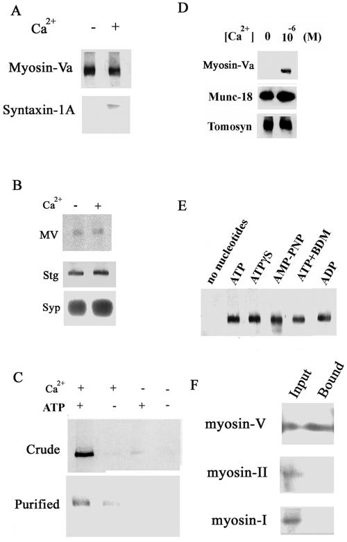
Ca2+-dependent binding of myosin-Va to syntaxin-1A. (A) Immunoprecipitation of the myosin-Va–syntaxin complex from brain homogenate using an anti-myosin-V antibody. The immunoprecipitation was carried out in the presence or absence of 10–6 MCa2+/2 mM Mg2+/0.5 mM ATP. (B) Myosin-Va (MV) is localized and remains on synaptic vesicles purified from cortex even after addition of 1 μM Ca2+. Myosin-Va was immunoprecipitated from brain homogenate in the presence and absence of 1 μM Ca2+, and myosin-Va, synaptotagmin I, and synaptophysin were detected by immunoblotting. Synaptotagmin I (Stg) and synaptophysin (Syp) are shown as synaptic vesicle markers. (C) Confirmation of the 10–6 M Ca2+/2 mM Mg2+/0.5 mM ATP-dependent binding of syntaxin-1A to myosin-Va in brain homogenate (Crude) or to purified myosin-Va (Purified). Binding of myosin-Va to GST-Syntaxin-1A was assessed in the presence and absence of Ca2+ and Mg-ATP using a GST pull-down assay, followed by anti-myosin-Va immunoblotting. (D) Ca2+-dependence of myosin-Va binding to syntaxin-1A. Brain homogenate was incubated with immobilized GST-syntaxin-1A in the presence or absence of 10–6 M Ca2+/2 mM Mg2+/0.5 mM ATP. Bound myosin-Va was detected by immunoblotting. Myosin-Va binding to syntaxin-1A required 10–6 M Ca2+. In contrast, binding of tomosyn and Munc-18 to syntaxin-1A does not require Ca2+. (E) Binding of myosin-Va by syntaxin-1A requires ATP analogues in presence of 10–6 M Ca2+. As shown by immunoblotting, myosin-Va bound to syntaxin-1A only in the presence of 0.5 mM ATP, ADP, or the nonhydrolyzable ATP analogues adenosine 5′-O-[3-thiotriphosphate] (ATPγS), 5′-adenylylimidodiphosphate (AMP-PNP), or 0.5 mM ATP with 5 mM 2,3-butanedione monoxime (BDM; a myosin-ATPase inhibitor). (F) Myosin-I (myr1A) and myosin-IIB do not bind to syntaxin-1A, even though they are present in the brain homogenate. Rat brain homogenate was mixed with mixed with GST-syntaxin-1A. Equal amounts (15 μl) of the rat brain homogenate (1 μg/μl; Input) and of the fraction bound to GST-syntaxin-1A (Bound; see C) were analyzed by immunoblotting with antibodies against myosin-Va, myosin-IIB (gift of T. Shirao, Gunma University Graduate School of Medicine, Maebashi, Gunma, Japan), or myr 1A (gift of M. Bähler, Westfalische Wilhelms University, Münster, Germany).

