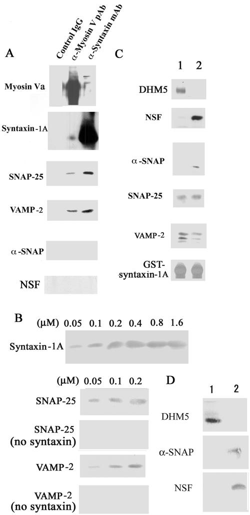
Figure 7.
Myosin-Va interacts with the SNARE complex via syntaxin-1A. (A) The myosin-Va–syntaxin-1A complex contains other SNAREs. The myosin-Va–syntaxin-1A complex was immunoprecipitated with an anti-syntaxin-1A antibody (α-syntaxin monoclonal antibody), an anti-myosin-Va antibody (α-Myosin-V pAb), or a control IgG from a pool of myosin-Va and syntaxin-1A-enriched fractions, which were obtained by 5–40% sucrose density gradient fractionation of hypotonically treated and Triton X-100-solubilized brain P2 fraction. The immunoprecipitated complexes were analyzed by immunoblotting. (B) Reconstitution study using recombinant SNAREs. Protein binding was assessed by immunoblotting. Top, His6-DHM5 (0.2 μM of dimer) immobilized on Ni2+-NTA resin was incubated with 0.05–1.6 μM of recombinant syntaxin-1A in the presence of 10–6 M Ca2+, and syntaxin-1A binding was assessed by immunoblotting DHM5 binding saturated at 0.2 μM syntaxin-1A. Bottom four, His6-DHM5-syntaxin-1A complex was formed with 0.05, 0.1, or 0.2 μM of syntaxin-1A and then incubated with the equal concentrations of SNAP-25 or VAMP-2. Controls contained no syntaxin-1A. Bound SNAP-25 and VAMP-2 were eluted with SDS-sample buffer and detected by immunoblotting. Note that SNAP-25 and VAMP-2 did not bind to myosin-Va in the absence of syntaxin-1A. (C) Reconstitution study using recombinant SNAREs, NSF, and α-SNAP. Immobilized GST-syntaxin-1A (0.2 μM) was mixed with (lane 1) or without (lane 2) DHM5 (0.2 μM dimer), and in the presence of 10–6 M Ca2+. NSF was incubated with α-SNAP for 2 h at 4°C to form a complex. This NSF–α-SNAP complex was mixed with VAMP-2 and SNAP-25 and then incubated for 2 h with GST-syntaxin-1A-DHM5 in the presence of 10–6 M Ca2+. Proteins bound to syntaxin-1A were visualized by immunoblotting. DHM5 bound to syntaxin-1A in the presence of SNAP-25 and VAMP-2 but not in the presence of NSF/α-SNAP. (D) The SNARE complex interacts with either α-SNAP/NSF or DHM5. The SNARE complex (0.2 μM) was formed as in B and C and then incubated for 1 h with α-SNAP/NSF (0.2 μM). This complex was then incubated for 1 h with DHM5 (1 μM) in the presence of 10–6 M Ca2+ (lane 1). Alternatively, DHM5 (0.2 μM) was first incubated with the SNARE complex followed by α-SNAP/NSF (1 μM) (lane 2). Bound proteins were detected by immunoblotting.

