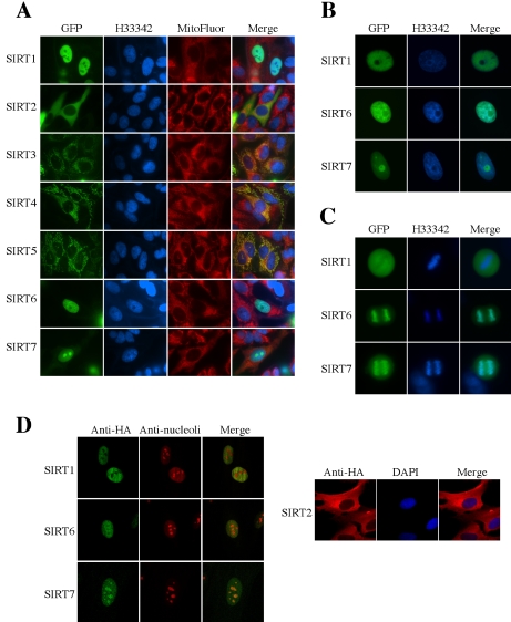
Figure 1.
Cellular localization of human SIRT proteins. (A) Each SIRT-GFP fusion protein was transiently expressed in NHF normal human fibroblasts. Hoechst 33342 (H33342) and MitoFluor stainings are shown in parallel. The merged pictures of GFP, Hoechst 33342 and MitoFluor are in the most right panels. (B) Magnified nuclei of NHF cells expressing SIRT1-GFP, SIRT6-GFP, or SIRT7-GFP protein. (C) Mitotic NHF cells expressing SIRT1-GFP, SIRT6-GFP, or SIRT7-GFP protein. Condensed chromosomes are strongly stained with Hoechst 33342 in the middle panels. (D) NHF cells expressing HA-tagged SIRT1, SIRT2, SIRT6, or SIRT7 were immunostained with anti-HA antibody, in combination with anti-nucleoli antibody (for SIRT1, SIRT6, and SIRT7) or DAPI staining (for SIRT2).

