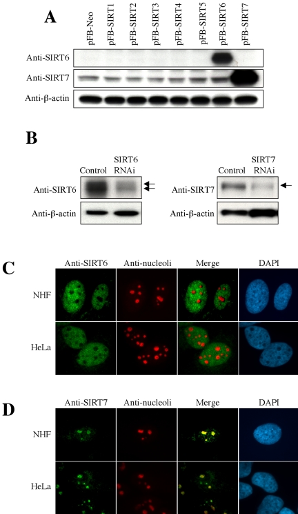
Figure 2.
Production of anti-SIRT6 and anti-SIRT7 antibodies and detection of endogenous SIRT6 and SIRT7 proteins. (A) NHF cells were infected with the retroviral vector driving each SIRT (pFB-SIRT1–7) or control vector (pFB-Neo) and used in Western blot with anti-SIRT6 and anti-SIRT7 rabbit polyclonal antibodies. β-actin was a loading control. (B) The endogenous SIRT6 (left) or SIRT7 (right) expression was knocked down by RNA interference (RNAi) in HeLa cells. The arrows indicate the specific signals, which were significantly weakened in the RNAi cells. The nature of two SIRT6 bands is currently unknown. (C and D) Indirect immunofluorescence staining of endogenous SIRT6 (C) and SIRT7 (D) proteins in NHF and HeLa cells. Costaining with anti-nucleoli antibody and DAPI staining are also shown.

