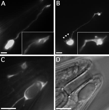
Figure 6.
Changes in the socket cell morphology in alr-1 mutant worms. (A–C) Fluorescence and (D) differential interference contrast images of unc53pB::GFP transgenic adults showing expression in the amphid socket cells. (A) Wild-type amphid socket cell morphology displaying the characteristic cylindrical distal ending (inset). (B) The amphid socket cell in alr-1(ok545) showing a typically misshapen distal ending (inset) and membrane projections near the cell body (triangles). (C and D) The distal end of an alr-1(ok545) amphid socket cell exhibiting a large intracellular vesicle. Anterior is to the upper right, posterior to the lower left. Bars, 10 μm.

