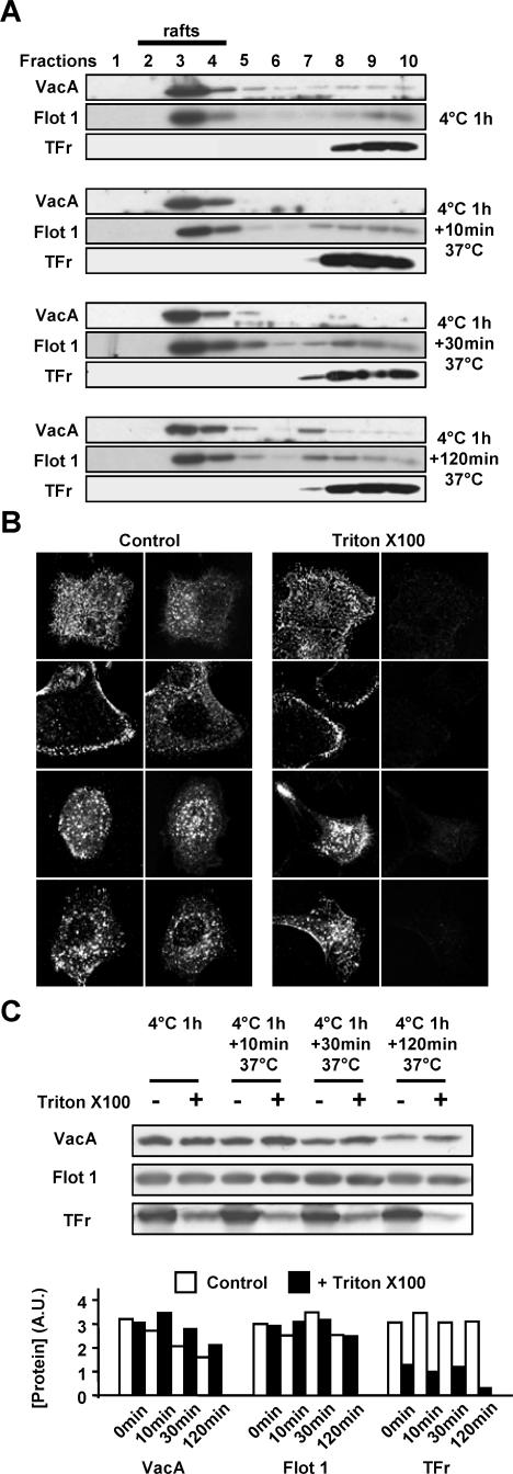
Figure 3.
Association of VacA with lipid rafts during endocytosis and intracellular trafficking. (A) AGS cells were incubated with VacA for 1 h at 4°C, washed and warmed for 0, 10, 30, or 120 min at 37°C. For each time point, cells were processed for lipid rafts analysis by flotation gradient after Triton X-100 extraction as described in Materials and Methods. Identification of the proteins in each fractions of the gradient was performed by immunoblots using the IgG 958 for VacA, the MAb anti-flotillin 1 for flotillin 1 (Flot 1; a marker of lipid rafts), and the MAb anti-TFr (as a protein marker excluded from lipid rafts). (B) VacA internalized in endocytic compartments is resistant to Triton X-100 extraction at 4°C by contrast to TFr. AGS cells were incubated with VacA at 4°C for 1 h, washed, and incubated for 0, 10, 30 or 120 min at 37°C. Cells were submitted or not to Triton X-100 extraction at 4°C (as described in Materials and Methods). Cells were fixed, processed for the detection of VacA and TFr by immunofluorescence, and observed by confocal microscopy. All pictures represent total cell reconstructions from confocal sections. Scale bar, 10 μm. (C) VacA, Flot 1, and TFr protein levels associated with control or Triton X-100-extracted AGS cells. The cells were treated as in B except that after Triton X-100 extraction, they were lysed and analyzed by immunoblots as in A. The histogram represents the quantification of the presented immunoblots (arbitrary units: A.U.).

