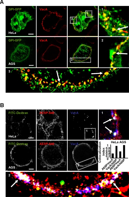
Figure 7.
VacA early endocytic compartments contain GPI-APs. (A) HeLa cells or AGS were transfected with GPI-GFP. Cells were incubated at 4°C and VacA was added for 1 h at 4°C. Cells were washed, incubated with VacA in warm medium for 10 min, and then processed for the detection of VacA (red) and GPI-GFP (green) by confocal microscopy. In zooms, arrows indicate the colocalization between VacA and GPI-GFP (yellow). (B) HeLa or AGS cells were incubated at 4°C for 1 h with the pan-GPI-APs marker ASSP-546 and VacA. Cells were washed and incubated in warm medium with FITC-dextran (4 mg/ml) for 10 min and processed for the detection of VacA (blue), ASSP-546 (red), and dextran (green). Arrows in the zooms show the colocalizations (white) between the markers. All pictures in A and B were taken from single confocal sections. Scale bars, 10 μm. The histogram represents the quantification of colocalizations between VacA and ASSP-546 by colocalization index. Results represent the account average for at least 12 cells for each column.

