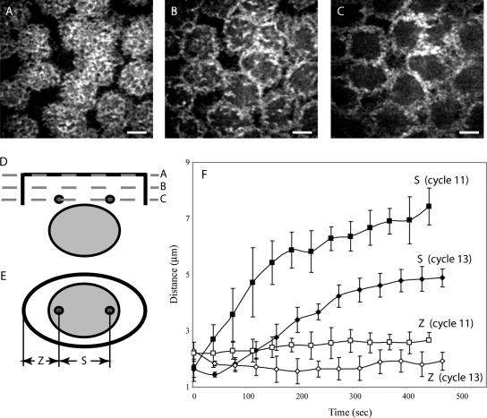
Figure 1.
Four-dimensional microscopy of a GFP-Polo-expressing embryo injected with rhodamine-actin. (A-C) Actin distribution in three consecutive confocal planes: at the cortex (A), 1 μm below the cortex (B), and 2 μm below the cortex (C). (D and E) Suggested geometry of the actin caps (plane A) and furrow (planes B and C) in relation to the position of the nucleus and centrosomes, shown in cross section, (D), and parallel to the cortex (E). (F) Pole-to-pole (S) and pole-to-furrow (Z) distances as functions of time in cycles 11 and 13. Bar, 5 μm.

