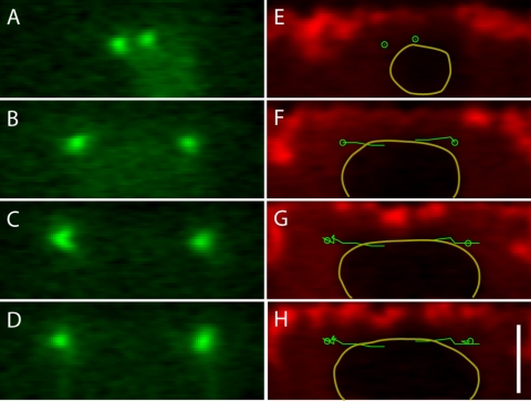
Figure 6.
Centrosomes separate along linear trajectories right under the cortex, whereas the nucleus deforms. Four frames from a reconstructed cross section of a time-lapse confocal stack (t = 0 s [A], 167 s [B], 503 s [C], and 615 s [D]) showing GFP-polo (A-D) and rhodamine-dextrans (E-H) with the positions of the centrosomes (green circles) and centrosome trajectories (green line) overlaid. The contour of the nucleus is outlined in E-H. Note that the apparent increase in the size of the nucleus is mostly due to the fact that the centrosomes and nucleus do not start off perfectly aligned so that a cross section of the stack through the centrosome at the beginning of separation cuts through the nucleus off-center. Bar, 5 μm.

