
Figure 1. Different quantitative imaging approaches.
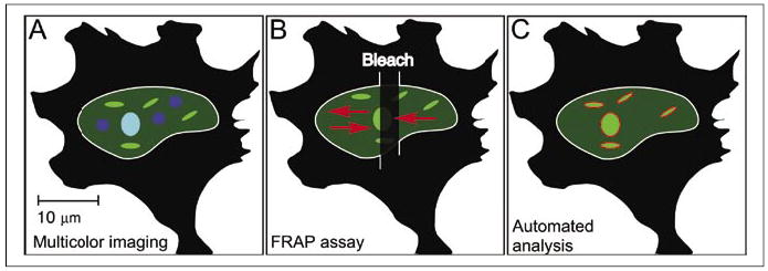
(A) Multicolor imaging to reveal protein co-localization can supply important information about the molecular composition of subnuclear domains. (B) The FRAP technique uses photobleaching of the labeled proteins within a ROI to measure the kinetics of the redistribution of the population of fluorescent-labeled proteins over space and time. (C) The analysis of subcellular structures using computer algorithms to automate the detection and measurement of subcellular features in large sets of high-resolution images. YFP, yellow fluorescent protein; NCoR, nuclear receptor corepressor protein; FRAP, fluorescence recovery after photobleaching; ROI, region of interest.