

Summary
The flowers of angiosperms are extraordinarily diverse. While most floral variation is visible to the naked eye, this diversity goes beyond the macroscale: Floral organs comprise an underappreciated range of cell types that generate a multitude of patterns across their surfaces and give rise to novel structures. Because diverse cell patterns provide adaptations to biotic and abiotic factors, they also contribute to angiosperm evolution and speciation. Yet, how such diversity originates remains to be understood. In this review, we focus on petals, which together form the corolla, to examine the mechanisms patterning floral surfaces at the cellular level. We summarize current research aiming to understand how cell fate specification and controlled cell growth (proliferation and expansion) are achieved with high spatial resolution during petal development. We also examine the adaptive potential for such patterns and how they contribute to plant fitness and diversification. Finally, we discuss promising directions for future research on the evolution of petal patterning at the microscale and identify outstanding questions that technological advances now make it possible to address.
Keywords: cell type, corolla, evo‐devo, floral development, natural variation, novelty, petal patterning, pollinator attraction
I. Introduction
Flowers – the reproductive structures of angiosperms – exhibit an extraordinary diversity of forms across the plant kingdom. A typical flower is comprised of male and female reproductive organs (stamens and carpel or pistil) surrounded by sterile parts: the petals, collectively known as the corolla, and an outer whorl of sepals, together forming the calyx (Endress, 2001). These structures are as functional as they are beautiful: A properly formed flower is vital for reproductive success of the plant. By facilitating self‐fertilization, wind pollination or pollinator attraction, floral morphology has a direct impact on reproduction and contributes to reproductive isolation and speciation (Barrett, 2010). As a result, the genetic mechanisms and adaptive significance of flower diversity provide an excellent framework for understanding the evolution and development of angiosperms.
Floral organs often vary in number, shape, symmetry or arrangement and the mechanisms accounting for the evolution of those traits have been extensively reviewed elsewhere (Litt & Kramer, 2010; Bowman et al., 2012; Bowman & Moyroud, 2024; Khojayori et al., 2024). However, many species produce highly specialized petals with complex shapes, lobed or fringed margins or the addition of various appendages. Such intricate petals are widespread, yet little is known about how these complex morphologies are created during development (Endress & Matthews, 2006).
Petal surfaces also harbour diversity at the microscopic scale, displaying a variety of cell types across their epidermis. The spatial distribution of different cell types is genetically encoded, allowing for the construction of robust patterns during development. As petal primordia emerge from the floral meristem, epidermal cells grow (proliferate and expand) and differentiate, acquiring specific colour, shapes and texture. Genetic modifications to petal cell behaviour during development yields morphological variation that can in turn contribute to the emergence of diverse patterns of cellular features or even specialized petal structures, such as nectar spurs (Moyroud & Glover, 2017). Therefore, exploring the mechanisms regulating cell behaviour across the corolla not only sheds light on the evolutionary processes accounting for the diversification of angiosperms, it also provides a framework to understand crosstalk between patterning and morphogenesis, two core developmental processes and their contribution to biodiversity.
In this review, we outline recent research on the mechanisms underlying the development of complex floral morphologies at the microscale, focusing on variation of petal traits between and within species that influence the evolution of flowering plants. Because petals constitute a major interface between a plant and its environment, cellular arrangements across the petal epidermis fulfil important adaptive functions, such as attracting pollinators and protecting reproductive organs in harsh environments. We highlight outstanding questions for understanding the mechanisms patterning the petal surface, and how emerging model organisms and novel technologies are being leveraged to understand petal evolution at the cellular level and revolutionize the field of petal evo‐devo. We discuss the genetic mechanisms that orchestrate cell differentiation to create patterns across the petal, by modifying epidermal cell features or specifying distinct cell types, and how these processes are selected upon to create novel patterns. We examine how the regulation of cell growth can be modified during development to create elaborate petal structures, such as nectar spurs, and how these principles can impact the evolution of novelty. Finally, we also outline recent studies that improve our understanding of petal patterns evolution in natural populations, arguing it is essential to investigate petal patterning across diverse species and accessions in the field.
II. Currently available model species provide diverse systems to understand petal evo‐devo at the microscale
Research in Arabidopsis thaliana largely established the basis of what is known about angiosperm development. However, A. thaliana depends predominantly on self‐pollination and its flowers are simple and small, making it difficult to answer questions about the evolution of complex flowers (Vallejo‐Marín & Barrett, 2009). As a result, model systems, such as Antirrhinum, Petunia and Mimulus (Fig. 1; Table 1), became foundational species for understanding petal development and evolution, and are now being used to study the role of flower morphology in natural selection and speciation (Schwarz‐Sommer et al., 2003; Yuan, 2019).
Fig. 1.

Model systems currently used to study flower evo‐devo at the microscale. (a) Brachypodium distachyon, (b) Phalaenopsis moth orchid, (c) Nigella sp., (d) Eschscholzia californica, (e) Aquilegia coerulea sp., (f) Delphinium sp., (g) Hibiscus trionum, (h) Clarkia amoena, (i) Mimulus guttatus, (j) Antirrhinum majus, (k) Petunia × hybrida and (l) Daucus carota. (m) Simplified phylogenetic tree of the angiosperms according to the APGIV classification (The Angiosperm Phylogeny Group et al., 2016), depicting the distribution of these model species. Pictures in (a, l) are credited to Laval University and Harry Rose, respectively, via Wikimedia Commons.
Table 1.
Examples of current model systems suitable to study flower evo‐devo at the microscale.
| Gene manipulation | Resources available | Petal cellular trait studied | References | Model system review | |
|---|---|---|---|---|---|
| Phalaenopsis (Moth orchid) | VIGS, stable transformation (tissue culture) | Genome, transcriptome, diverse species and accessions | Pigmentation, cell shape, cuticle texture, scent, organ and elaboration |
Lu et al. (2022) Hsu et al. (2021) Liao et al. (2020) Pramanik et al. (2020) Mao et al. (2024) |
Tsai et al. (2008) Zhang et al. (2023) |
| Nigella | VIGS | Chloroplast genome, transcriptome and inter‐ and intraspecies variation | Pigmentation, pseudonectaries and cell shape |
Yuan et al. (2023) Yao et al. (2019) Liao et al. (2020) Zhang et al. (2020) |
Damerval & Becker (2017) |
| Eschscholzia (California poppy) | VIGS | Draft genome and transcriptome | Pigmentation and cell shape |
Wilts et al. (2018) Pollack et al. (2019) |
Becker et al. (2023) |
| Aquilegia (columbine) | VIGS | Genome, transcriptome, inter‐ and intraspecific variation and mutant collection | Nectar spur and nectary development |
Cabin et al. (2022) Edwards et al. (2022) |
Kramer (2009) Kramer & Hodges (2010) Yant et al. (2015) von Balthazar et al. (2025) |
| Delphinium | VIGS | Chloroplast genome, transcriptome and natural interspecific variation | Organ morphology, cell shape, pigment and nectar spur development | Zhang et al. (2024) | Samarah et al. (2020) |
| Hibiscus | Stable transformation (tissue culture) | Annotated genome, transcriptome, inter‐ and intraspecific variation and CRISPR/Cas9 gene editing | Pigmentation, cuticle texture and cell shape |
Moyroud et al. (2022) Riglet et al. (2024) Yao et al. (2019) |
|
| Clarkia | VIGS | Genome draft and natural intraspecific variation | Pigmentation and scent | ||
| Mimulus (monkeyflower) | Stable transformation (floral dipping) | Annotated genome, transcriptomes, collection of mutants and natural accessions, inter‐ and intraspecific variation and CRISPR/Cas9 gene editing | Pigmentation (biosynthesis of pigments and complex pattern formation), corolla tube formation and flower symmetry |
Liang et al. (2023) Gao et al. (2024) Zhang et al. (2021) |
Yuan (2019) |
| Antirrhinum (snapdragon) | NA | Genome, transcriptomes, mutant collection, inter‐ and intraspecific variation and horticultural varieties | Pigmentation and cell shape |
Durán‐Castillo et al. (2021) Tavares et al. (2018) |
Davies et al. (2018) Schwarz‐Sommer et al. (2003) Mizzotti et al. (2014) |
| Petunia | Stable transformation (tissue culture) | Genome, transcriptome, mutant collection, inter‐ and intraspecific variation, horticultural varieties and CRISPR/Cas9 gene editing | Pigmentation (visible & UV), cell shape and petal cuticle synthesis |
Chopy et al. (2023) Skaliter et al. (2023) Li et al. (2022) |
Vandenbussche et al. (2016) Strazzer et al. (2023) |
| Daucus carota (wild carrot) | Stable transformation (tissue culture) | Genome, transcriptome, natural accessions and CRISPR/Cas9 gene editing | Pigmentation and complex floral architecture |
Duan et al. (2024) Baczyński et al. (2022) |
Que et al. (2019) |
| Brachypodium distachyon | Stable transformation (tissue culture) | Annotated genome, transcriptome, collection of mutants and natural accessions, inbred lines and CRISPR/Cas9 gene editing | Lodicule, lemma and glume development | Patterson et al. (2024) |
Schrager‐Lavelle et al. (2017) Raissig & Woods (2022) Kellogg (2015) |
VIGS, Virus‐Induced Gene Silencing.
The establishment of an increasingly expanding range of diverse model organisms (Fig. 1; Table 1) is allowing for the transition from descriptive studies of floral patterns to more mechanistic understanding of complex pattern modification and the evolutionary consequences of such changes. Species recently developed as models with more complex floral structures, including Aquilegia, Hibiscus and Nigella, open new directions by helping to understand how and why elaborate corollas evolve (Kramer & Hodges, 2010; Zhang et al., 2020; Cabin et al., 2022; Raissig & Woods, 2022; Cheng et al., 2023; Yuan et al., 2023; Riglet et al., 2024). Monocot models also offer promising research avenues. According to the floral quartet model, members of the MADS‐box gene family code for transcription factors that combine with each other to form distinct tetrameric complexes (MADS‐box complexes) that specify different floral organ identities (Theissen & Saedler, 2001; Bowman & Moyroud, 2024). Interestingly, recent functional investigations in orchids have uncovered that distinct MADS‐box complexes also specify sophisticated morphologies and pigmentation patterns across the perianth of Phalaenopsis species (Hsu et al., 2015, 2021; Pramanik et al., 2020). Grasses lack petals but have analogous organs called lodicules and genetic models, such as Brachypodium, are attractive systems to investigate whether those species deploy similar programmes to eudicots to pattern the surface of their floral organs (Schrager‐Lavelle et al., 2017). As transformation protocols are developed for an increasing number of organisms, we can now dissect the cellular mechanisms of more complex petal patterns and elucidate the genetic basis of floral traits that are not found in traditional models.
Additionally, due to the decreasing cost of whole‐genome sequencing and de novo assembly, studies using population genomics and genome scans are becoming more feasible in nonmodel systems. This is allowing for a number of recent studies that take advantage of naturally occurring genetic variation in situ to understand population genomics, hybridization and natural selection as it occurs in real time (Bradley et al., 2025; Field et al., 2025; Richardson et al., 2025). These resources can also assist in mapping complex quantitative traits with fine resolution, using quantitative trait locus (QTL) mapping and genome‐wide association studies. For example, a study in sunflowers used natural accessions to dissect the genetic basis of ultraviolet (UV) pattern diversity (Todesco et al., 2022). Together, these highlight a need for the collection of accessions in other species to encompass a wide genetic and phenotypic diversity at the cellular level. Such resources would likely illuminate petal evo‐devo at the microscale, echoing the contributions the 1001 Genomes project has made to our understanding of adaptation and evolution in A. thaliana (Alonso‐Blanco et al., 2016).
III. Cell patterns on the petal surface as adaptive traits
Patterns created on the petal epidermis are ecologically relevant because they mediate biotic and abiotic interactions. For example, corolla appearance is important for attracting pollinating animals and has long been credited for the rapid diversification of angiosperms as it can lead to reproductive isolation and speciation (Van der Niet et al., 2014). A number of recent studies have begun to reveal novel functions for these patterns and markedly advance our understanding of their biological significance.
Petal epidermal patterns are best known for their role in pollinator attraction. These patterns can enhance visibility of a flower from a distance or act as guides to direct insects to nectar or pollen after landing (Free, 1970; Whitney & Glover, 2007; Ojeda et al., 2016; Costa et al., 2017). Some of these motifs are not always perceptible by the human eye, such as patterns in UV‐absorbing or reflecting properties across the petal that often correlate with local pollinator vision (Fig. 2). For example, the gain of UV reflection in the petals of red poppies as they spread across Europe coincides with a shift in pollination system from beetles to bees (Martínez‐Harms et al., 2020). The presence of UV patterns on the corolla of many red flowers in the Mediterranean basin is proposed to be adaptations for pollination by hymenopterans (red‐blind insects) in environments where bird‐mediated pollination is not present (León‐Osper & Narbona, 2022). Motifs on the corolla can also lure pollinators by resembling food supply (Ma et al., 2016; Lunau et al., 2024) or brooding sites by mimicking the fungi fruiting bodies on which some species of flies lay their eggs (de Melo et al., 2011; Johnson & Schiestl, 2016; Abrahamczyk et al., 2021). In orchids and daisies, petal patterns also participate in sexual deception by emulating a pollinator's mating partners (Ellis & Johnson, 2009; Gaskett, 2011).
Fig. 2.
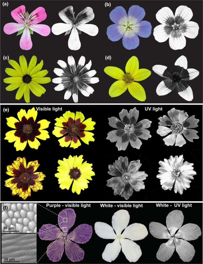
Differences in ultraviolet (UV) light reflecting and absorbing pigments as well as cell shape and texture across the petal surface can generate petal patterns in the UV range. (a–d) Images of flowers in human‐visible and UV light spectrums of (a) Pelargonium ‘Pink capitatum’ (Geraniaceae), (b) Geranium sylvaticum (Wood Cranes‐Bill, Geraniaceae), (c) Rudbeckia laciniata (Cut‐leaf coneflower, Asteraceae) and (d) Bidens triplinervia (Asteraceae), each with variation in UV‐absorbing or reflective properties across the corolla. (e) Natural variation in petal pigmentation patterns and UV‐reflective properties of Coreopsis tunctoria (Nuttall Weed, Asteraceae), with variation in UV reflection varying with both pigment and cell texture. (f) Artificial epoxy resin flowers created by imprinting the petal of Hibiscus trionum in dental wax to replicate its surface: striated flat elongated cells in the proximal portion and smooth conical cells in the distal portion of the petal, as seen under SEM imaging (Top left: SEM image of distal smooth conical cells; Bottom left; SEM image of proximal flat striated cells). In the purple artificial flower (left), the conical cells create a mat velvety appearance on the corolla periphery while the flat striated cells render its centre shiny. This ‘structural’ bullseye is not easily visible to the human eye in the white artificial flower (middle) but become apparent when imaged with a UV camera (right), showing that pigment, shape and texture of epidermal cells all influence visible and UV light reflective properties of the petal.
Petal patterns can also protect reproductive organs from stress, including heat, UV exposure, drought and herbivory. Larger UV bullseye patterns in silverweed and sunflower populations are believed to protect pollen from damage in environments with high levels of UV exposure (high altitude, artic and hot climates) or allow flowers to better resist desiccation (Koski & Ashman, 2015; Koski et al., 2020; Todesco et al., 2022). However, such a role might be species‐specific, as populations of Clarkia unguiculata exposed to lower levels of solar radiation produce larger, not smaller, UV‐absorbing bullseyes, suggesting that they do not provide significant pollen protection against UV rays (Peach et al., 2020). This advocates for similar studies to be conducted in a broader range of species to try and identify general trends, if any exist.
IV. Organizing and modifying the colour, shape and texture of petal pavement cells
Patterns emerge across the petal surface when pavement cells in distinct regions of the epidermis display contrasting characteristics: Petal epidermal cells can vary in shape, texture of their waxy cuticle and pigment content (Fig. 3). Recent studies have begun to identify the mechanisms involved in programming and modifying pavement cell properties across the corolla.
Fig. 3.

Micropatterns on the petal surface are created by variation in epidermal cell pigment, shape and cuticle textures. (a) Patterns on the petal of Erodium castellanum (Geraniaceae) created by cell‐specific pigmentation. (b) Variation in cell shape across the petals of Hardenbergia violaceae (Fabaceae). (c) Smooth cuticle on elongated petal epidermal cells of Ceratostigma griffithii (Plumbaginaceae). (d) Wrinkled cuticle on the elongated petal epidermal cells of Gaura lindheimeri (Onagraceae). (e) Smooth cuticle on the conical petal epidermal cells of Hibiscus trionum (Malvaceae). (f) Wrinkled cuticle on the conical petal epidermal cells of Dahlia merckii (Asteraceae).
1. Spatial control of petal pigmentation
Pigmented patterning on the petal surface is most well studied of these epidermal cell characteristics. The main classes of compounds that colour the corolla are flavonoids (red to blue anthocyanins, yellow chalcones and aurones or UV‐absorbing flavonols), carotenoids (reds, orange and yellows), betalaïns (reds and blues, exclusive to the Caryophyllales) and chlorophylls (greens), and their biosynthesis pathways are well understood (Tanaka et al., 2008; Tripathy & Pattanayak, 2012). The chemical properties of these molecules are highly modifiable, such as the addition/removal of sugar or methyl moieties or changes in pH, which allows for exceptional variation in pigmentation across flowers (Morita et al., 2015; Stavenga et al., 2021). Colourful petal patterns are created by the precise production of these compounds in different epidermal cells across the petal surface (Fig. 3a; Fattorini et al., 2024; Lin & Rausher, 2021b; Yuan et al., 2023). Occasionally, pigments in underlying mesophyll cells can also contribute to the overall effect of pigmentation patterns (Cavallini‐Speisser et al., 2021; Yuan et al., 2023).
Evolving intricate pigmentation patterns, such as scattered speckles, eye‐like concentric spots or juxtaposed horizontal stripes, requires the genetic machinery that produces different pigments to be spatiotemporally restricted during development. This process is often regulated by the myeloblastosis (MYB)‐domain family of transcription factors (Zhao et al., 2022). For example, petal anthocyanin biosynthesis is regulated by MYB transcription factors as part of a trimeric complex also involving a bHLH TF and a WD40 protein (MBW complex) that transcriptionally activates different pigment biosynthetic genes (Xu et al., 2015). MYB‐bHLH‐WD complexes also regulate carotenoid and betalaïn biosynthetic processes in many species, suggesting that these pathways are evolutionarily conserved (Hatlestad et al., 2015; Sagawa et al., 2016; Lloyd et al., 2017; Zhao et al., 2022). The spatial restriction of MYB genes to specific subdomains of the emerging petal primordia often accounts for the formation of pigmentation patterns (Fig. 4a–c). Variation in the expression pattern of MYBs and their co‐activators accounts for the majority of flower colour variation across and between species (Fig. 4d–f), as surveys of genetic variation in flower pigmentation across species found that the genes encoding the MBW complexes themselves display faster rates of molecular evolution than their downstream biosynthetic enzymes (Schwinn et al., 2006; Wessinger & Rausher, 2012; Wheeler et al., 2021, 2022). This phenomenon is likely adaptive, as it allows for the preservation of pigment biosynthesis pathways for other processes, such as stress responses and the pigmentation of other organs (Davies et al., 2018).
Fig. 4.

MYB transcription factors are major contributors to petal pattern development and evolution. (a) Spatial restriction of MYB gene expression (e.g. subgroup 6 (SG6) R2R3‐myeloblastosis (MYB) promote anthocyanin production while subgroup 7 (SG7) R2R3‐MYB activate flavonol synthesis) largely accounts for pigmentation pattern formation during petal development; (b) variation in expression domain or (c) in the identity of the MYB gene expressed in a given region (subgroup 21 R2R3‐MYB can promote the production of yellow carotenoids) can generate the intra‐ and interspecific diversity in petal patterns. (d) Variation in pattern proportions can be due to mutation affecting the coding sequence or the regulatory region of MYB genes. Red crosses depict deleterious mutations, and green crosses represent gain of cis‐regulatory elements. (e) Petal pigmentation spots can emerge from the interactions between a transcriptional activator (SG6 R2R3‐MYB) and a repressor (e.g. R3‐MYB). Green arrows indicate transcriptional activation, red blunt arrow indicate transcriptional repression. (f) Post‐transcriptional processes targeting SG6 R2R3‐MYBs can also yield petal pattern variation, reducing pigment production via changes in the 5′UTR impacting protein translation (right) or abolishing pigment production in certain petal domains via siRNA promoting the degradation of SG6 R2R3‐MYB transcripts (left). Arrows indicate transcription and translation steps. (g) MYB genes are also involved in specifying cell shape and cuticular texture, but many outstanding questions remain: Beyond bHLH/WD40 that help MYB regulate anthocyanin production and AP2/ERF factors that participate in cuticular ridges formation, most of the molecular players working along MYBs to control epidermis cell characteristics are yet to be identified. What specifies the geometry of nonconical cell shape and what decides whether SG9 MYBs induce conical cell or trichome formation is also not understood. (h) Crucially, the signalling events and upstream regulators that specify petal polarity, pattern cell fate along the axes of developing petal primordia and divide the petal surface into distinct territories where growth can be controlled independently, and neighbouring cells can acquire contrasting fates remain unclear. Brown arrows indicate the developmental progression from emerging primordia on the floral meristem to mature petals in open flowers.
Diversification of pigmentation patterns due to evolutionary drivers are mediated by changes in the composition, type and relative position of the different elements of a pattern. From a theoretical viewpoint, petal pigmentation patterns can be formed and diversify following mechanisms similar to those responsible for colourful motifs on the skin or fur of animals (Galipot et al., 2021). Repetitive patterns like small, scattered spots can emerge via self‐organization following the Turing model and only require interactions between a transcriptional activator and a repressor (Turing, 1990; Meinhardt, 2012). In this model, expression of the inhibitor is promoted by the activator. The inhibitor then represses the pigmentation activator in nearby tissue, creating spots or stripes across an organ (Fig. 4e; Galipot et al., 2021).
The Turing reaction–diffusion model has been validated both in silico and in vivo in Mimulus (Fig. 1i). Red spots on the nectar guide of the yellow petals of M. lewisii are created by synchrony between RTO, a MYB repressor of anthocyanin production, and NEGAN, a subgroup 6 R2R3‐MYB that activates anthocyanin production. In red‐spotted regions, NEGAN promotes anthocyanin biosynthesis and RTO expression. An active RTO protein can move into adjacent tissues to repress NEGAN expression and anthocyanin production, maintaining yellow tissue around the red spot (Fig. 4e). This phenotype is likely also adaptive, as a loss‐of‐function mutation in RTO produces a ‘red‐tongue’ phenotype, in which the discrete spots are replaced with a large blotch of colour, resembling wild Mimulus populations across California (Ding et al., 2020).
Computational approaches to model petal development have been extremely useful in understanding the emergence of distinct corolla patterns (Rolland‐Lagan et al., 2003; Green et al., 2010; Sauret‐Güeto et al., 2013; Rebocho et al., 2017; Zhang et al., 2024). More recently, theoretical models have been developed specifically to better understand the processes that led to the emergence of various pigmentation patterns (Ringham et al., 2021; Simmons et al., 2023; Liang et al., 2025). Genes do not work in isolation but instead interact with each other, forming information processing webs, commonly known as gene regulatory networks (GRNs), that control cell behaviour, such as differentiation and growth. Novel in silico simulations of developing petals at cellular resolution should also allow us to investigate how the GRNs that govern petal patterning originate and how complex patterns evolve (Rolland‐Lagan et al., 2003; Ringham et al., 2021; Zhang et al., 2024). Early attempts are promising: They demonstrate how the spontaneous specification of an additional boundary cell type can contribute to the emergence of bullseye patterns, uncover the importance of noise in GRN to generate robust petal motifs and establish growth as a central player to maintain or modify pattern proportions as petals develop (Riglet et al., 2024; Oud et al., 2025).
Recent gene duplication events play a central role in the diversification of petal pigmentation patterns, allowing closely related MYBs to acquire distinct expression patterns and/or biochemical properties. This accounts for the production of the complex eyebrow‐like horizontal stripes of Nigella orientalis (Yuan et al., 2023) and the formation of red spots and white cups on the petals of Clarkia gracilis (Lin & Rausher, 2021b). Differences in the positioning of red petal spots and relative size of the white cup domain between subspecies of C. gracilis are also due to loss‐of‐function mutations, gene loss and cis‐regulatory changes affecting the same MYB paralogues (Fig. 4d; Lin & Rausher, 2021a).
MYBs can also regulate UV patterns. For example, sunflowers and many other Asteraceae have UV bullseye patterns created by the spatial restriction of UV‐absorbing flavonoids to the proximal region of the corolla in ray florets, the peripheral flowers of the Asteraceae inflorescence (Moyers et al., 2017; Fig. 2c–e). A study in wild sunflowers uncovered that intraspecific differences in UV‐absorbing bullseye are controlled by cis‐regulatory variation in a MYB gene (Fig. 4d; Todesco et al., 2022). This work, along with other recent investigations (Koski & Ashman, 2015; Bradley et al., 2017; Ding et al., 2020), underscores that natural variation is a powerful tool to investigate the genetic basis of petal pattern diversification. It also provides a path to identify the upstream processes regulating MYB expression, as polymorphisms in the promoter region of HaMYB111 that associate with bullseye dimensions could help identify the binding sites of upstream transcription factors.
While changes to gene transcription have been the dogma for the evolution of petal pigmentation, new studies indicate that the regulation of complex patterns also rely on posttranscriptional mechanisms (Fig. 4e). In Mimulus lewisii, the YELLOW UPPER (YUP) locus produces siRNAs that silence REDUCED CAROTENOID PIGMENTATION2 (RCP2), a MYB regulator of carotenoid production (Liang et al., 2023). Variation in the YUP locus accounts for the production of a yellow nectar guide on the pink corolla of the bee‐pollinated M. lewisii or the uniform red pigmentation of the bird‐pollinated M. cardinalis and swapping YUP alleles between species is sufficient to modify pollinator preference (Bradshaw & Schemske, 2003). Together, this suggests that this small RNA locus played a key role in diversification and pollinator‐mediated reproductive isolation in closely related Mimulus species (Peng et al., 2024).
These findings complement earlier research in wild populations of snapdragon (Antirrhinum majus) subspecies Antirrhinum majus subsp. striatum and Antirrhinum majus subsp. pseudomajus (Whibley et al., 2006). Researchers found a small RNA named SULF that targets chalcone 4′‐O‐glucosyltransferase (Am4′CGT), a gene that is important in synthesis of the yellow pigment (Bradley et al., 2017). Am4′CGT and SULF expression patterns are complementary, suggesting that SULF posttranscriptionally represses Am4′CGT. SULF is also epistatic to a MYB‐domain transcription factor regulating red pigment synthesis, ROSEA (ROS), which highlights complex mechanisms for regulating different pigments across the petal (Whibley et al., 2006; Bradley et al., 2017). In addition, SULF is under selection in naturally hybridizing populations of A. majus subsp. striatum and A. majus subsp. pseudomajus, indicating this important in the evolution of petal patterning in Antirrhinum.
Interestingly, both of SULF and YUP are taxa‐specific and were a result of by incomplete or inverted duplication of protein‐coding genes. In Mimulus, the partially copied gene is a CYP450 homologue that is unrelated to pigment production (Liang et al., 2023). Together, these studies highlight the role of posttranscriptional processes in the evolution of complex pigmentation patterns – it should also encourage researchers mapping quantitative trait loci in other model systems to look beyond protein‐coding genes.
2. Patterning cell shape across the petal epidermis
Epidermal cells can also vary in size, shape and texture across the petal (Fig. 3b–f; Christensen & Hansen, 1998; Kay et al., 1981). The distribution of these structural characteristics is controlled with remarkable precision: The petal surface of Nigella arvensis involves the arrangement of more than eight different cell shapes (Yao et al., 2019). Petal cell shape can also vary extensively between species and different petals of zygomorphic flowers often exhibit distinct cell geometries patterns (Bailes et al., 2018; Liao et al., 2020).
Variation in cell shape across the petal likely has an adaptive role and can impact pollinator preference. For example, in isolated populations on the Macaronesian islands, the loss of conical cell shape on the petal epidermis has occurred independently at least five times in eudicots species that have shifted from primarily insect to bird pollination (Ojeda et al., 2016). However, a more recent comparative study of species‐pairs across 13 genera (11 asterids and 2 rosids) found no correlation between petal epidermal cell shape or texture and pollination mechanism (Kraaij & van der Kooi, 2020). This suggests the ecological relevance of petal epidermal cell shape variation may differ between species and emphasizes the need for more detailed characterization of the contribution of cell shape to angiosperm evolution.
The most well‐studied petal epidermal cell shape is a dome‐like cone (Fig. 3e,f). These conical cells are present on the petals of most species, often found combined with other cell shapes (Kay et al., 1981). Conical cells contribute to pollinator attraction by enhancing the colour of the flower (Noda et al., 1994) and are also important for physical properties of the petal, including preventing petal wettability, regulating flower temperature and providing grip for pollinating insects (Whitney et al., 2009). Subgroup 9 MYB transcription factors, also known as MIXTA and MIXTA‐like genes, regulate conical cell formation (Fig. 4g; Brockington et al., 2013; Reed et al., 2022). MIXTA‐like genes are widely conserved as regulators of epidermal cell shape across angiosperms and variation in conical cell distribution across the petal correlates with their expression pattern during development. For example in Thalictrum, the loss of conical cell shape corresponds with changes in expression of a MIXTA‐like gene and is correlated with the transition from insect to wind pollination (Di Stilio et al., 2009). However, the identity of the transcriptional regulators specifying other petal epidermal cell shapes is still mysterious (Fig. 4g). Overall, the GRNs controlling how and where distinct cellular geometries emerge in a robust manner across the corolla are still largely unknown and are important future research venues (Cavallini‐Speisser et al., 2021; Riglet et al., 2021).
3. Cell texture: Variation in cuticle properties across the petal epidermis
A third, less appreciated element of petal epidermal patterning is the cuticle texture. A wide range of cuticular patterns exists across the petals of angiosperms, and research over the last few years has started to highlight their contribution to corolla diversification (Fig. 3c–f). The cuticle texture can vary across a single petal (Moyroud et al., 2022) and contributes to the corolla appearance: Disordered striations created by alternate ridges and grooves on the cuticle of petal epidermal cells contribute to structural colour and UV patterns yielding visual cues to pollinators (Moyroud et al., 2017). Cuticular patterns also act as water repellent, influence temperature on the corolla surface and impact attachment and flower handling by visitors (Koch et al., 2009; Bräuer et al., 2017; Wilts et al., 2018; Gorb & Gorb, 2023). Cuticle structure likely influences floral scent emission and produces the ‘velvety’ and ‘waxy’ textures of orchid petals that prevent insect adhesion (Prüm et al., 2011; Xiao et al., 2020; Liao et al., 2021).
Theoretical models proposed that the rate of cuticle production, the extent and direction of cell expansion, the relative stiffness of the cuticle and the underlying cell wall properties are all important to control the production, direction and regularity of ridges on petal epidermal cells (Antoniou Kourounioti et al., 2013; Airoldi et al., 2021; Lugo et al., 2023). The chemical composition of the cuticle also influences its material properties and varies between species (Kunst & Samuels, 2009). For example, the presence of cuticular striations in flowers belonging to a group of Hibiscus species native to Australia and New‐Zealand (Fig. 2f) is best predicted by examining the accumulation of waxes and phenolic compounds in the petal epidermis cuticle (Moyroud et al., 2022).
SHINE transcription factors from the AP2/ERF family were first identified as regulators of cuticle ridge formation in A. thaliana petals (Fig. 4g; Shi et al., 2011; Oshima et al., 2013). Work done in Hibiscus trionum suggests that gene duplication followed by functional divergence could be central to the emergence of different cuticular patterns across the petal. H. trionum petals exhibit flat striated cells in the proximal portion and smooth conical cells in the top region, with different cuticle chemical profiles between the two domains (Fig. 2f; Giorio et al., 2015). While HtSHINE1 and HtSHINE2 are lowly expressed across the emerging petal, HtSHINE3 is highly transcribed only in the striated domain and can modify cuticle composition and induce ectopic striations when constitutively overexpressed (Moyroud et al., 2022).
MYB‐domain transcription factors also participate in the control of cuticle production and ridges formation (Fig. 4g; Xu et al., 2021). In A. thaliana, MIXTA‐like transcription factors, together with SHINE genes, act redundantly to induce conical cell formation and striation emergence (Oshima et al., 2013; Shi et al., 2011), providing a simple way to co‐regulate both cell shape and texture. However, this is not conserved across all species, as HtMIXTA‐like1 regulates cuticle composition and striation production but does not participate in the specification of the conical cell shape on H. trionum petals (Moyroud et al., 2022).
4. Petal pavement cell features work in combination
Although a systematic investigation across the angiosperms is lacking, targeted studies indicate that changes in epidermal cellular features often occur in combination with each other, creating petal subdomains with distinct properties (Berry & Geeta, 2019; Riglet et al., 2021; Berry et al., 2023). For example, floral UV patterns can be created through variation in cell pigments, shapes and cuticle texture, or a combination of the three (Fig. 2a–f). UV‐absorbing pigments (flavonoids, carotenoids but also other compounds like lignin) have long been known to produce motifs on the corolla. However, more recent studies revealed that structural characteristics of epidermal cells also matter: cuticular striations, when associated with flat elongated cells, can act as semi‐ordered diffraction gratings, producing blue/UV‐reflective cues that pollinators can perceive (Moyroud et al., 2017). The cell architecture of the petal surface can also enhance UV absorption on its own, as replicas of corolla surface using transparent resin still produce UV patterns mimicking those present on the surface of real flowers (Fig. 2f; Schulte et al., 2019).
Petal cell pigmentation, shape and texture can work together to mediate evolutionary innovations. The Beetle daisy (Gorteria diffusa) exhibits a remarkable range of intraspecific variation, with different floral morphs found in geographically distinct populations (Ellis & Johnson, 2009; Thomas et al., 2009). Some populations display raised black spots on petal‐like ray florets that resemble the primary pollinator of this species, a small bee‐fly called Megapalpus nitidus. These spots are a form of visual mimicry, as they resemble female bee‐flies that attract males pollinators through sexual deception (Johnson & Midgley, 1997). Such elaborate tridimensional structures are created by localized changes in epidermal cell pigment, shape, texture and UV reflectance, each contributing to the complex appearance of petal spots (Johnson & Midgley, 1997; Thomas et al., 2009; Fattorini et al., 2024).
Surprisingly, the genetic pathways associated with petal spots in Beetle daisy participate in iron homeostasis, root hair production and control of the juvenile to adult vegetative transition (Kellenberger et al., 2023). These gene networks contribute to unrelated processes may have been sequentially co‐opted to facilitate the creation of a phenotypic novelty and take advantage of insect mating behaviour to favour pollination. For example, the miR156/SPL module is known to be important early in vegetative development, but a role in specifying petal spots, where miR156 was not known to be highly expressed, is an exciting novel function for the evolution of this pathway (Poethig & Fouracre, 2024). While the molecular mechanism by which these developmental pathways are repurposed to create these spots is not yet understood, it provides an interesting example of how evolution can drastically alter and combine different epidermal cell traits to create diversity on the petal surface. The development of protocols to genetically manipulate Beetle daisy will be a promising venue to test the hypothesis that combined gene co‐options enabled the rapid evolution of a novel and complex trait. It would also be interesting to know whether other unrelated South African species of Gazania, Dimorphotheca and Pelargonium that display similar dark spots (Johnson & Midgley, 1997) evolved these patterns using the same genetic pathways.
5. Beyond MYBs: revealing the upstream mechanisms patterning the petal epidermis
Little is known about the upstream regulators of MYBs associated with petal pigmentation and the nature of their contribution, if any, to the diversification of colour patterns (Fig. 4h). Recent research has uncovered some candidate transcription factors which could regulate the expression of the MBW complex: In the Beetle daisy (Gorteria diffusa), presence of a pigmented black spot on petals coincides with high expression of a GdSPL1, a SQUAMOSA PROMOTER BINDING PROTEIN‐LIKE transcription factor. Interestingly GdSPL1 is silenced in unspotted petals by the microRNA, miR156 and co‐option of the miR156‐SPL module (a gene network involved in juvenile to adult leaf development) could have been instrumental in establishing spot placement (Kellenberger et al., 2023). However, whether GdSPL1 promotes anthocyanin production and acts as an upstream regulator of the MBW complex to create the darkly pigmented spot characteristic of this species remains to be tested. Similarly, little is known about the upstream processes responsible for the spatiotemporal control of MIXTA and MIXTA‐like genes expression to restrict conical cell production or the formation of cuticular striations to certain petal domains (Fig. 4g). In Lotus japonicus, a CYCLOIDEA homologue, LjCYC2, activates the expression of subgroup 9 MYBs to restrict conical cell production to the very base of wing petals (Ojeda et al., 2017). Another CYC homologue, TfCYC2 in Torenia fournieri, was shown to directly bind to the promoter of TfMYB1, a regulator of pigment production in this species, to promote its expression during petal development (Su et al., 2017). However, whether CYC genes act upstream of MYBs to regulate cell shape or pigmentation in other species has not been examined.
More generally, the upstream regulatory events that restrict the expression of MYBs and other transcription factors that control pigment synthesis, cell shape or cuticular texture to specific domains of the corolla remain largely obscure, but new research suggests these patterns are defined very early in development (Fig. 4h). Recent data in Hibiscus indicate that the petal epidermis is divided into distinct subdomains when developmental boundaries are set long before any visible pattern emerges on its surface (Riglet et al., 2024). This creates a ‘paint‐by‐number’ canvas, allowing cell behaviour to be controlled independently in different subregions. Importantly, variation in the positioning of the prepattern boundary coincides with a change in bullseye dimension between H. trionum and its sister‐species H. richardsonii. Changes in pattern proportions could have contributed to speciation in these two species, as buff‐tailed bumblebees exhibit a strong preference for the larger bullseye of H. trionum over the smaller pattern of H. richardsonii (Riglet et al., 2024). However, the molecular players, signalling processes and cellular events involved in partitioning the corolla so early in development are still unknown. Signalling events that specify petal polarity are likely to play a key role in patterning cell fate specification and growth across the surface of developing petal primordia (Cavallini‐Speisser et al., 2021). Hormones, like auxin, are obvious candidates (Salvi & Moyroud, 2025), but other molecular players like small peptides or secondary metabolites should also be considered (Fig. 4h).
High spatial resolution approaches, such as single‐cell or single‐nuclei RNA‐seq, should greatly enhance our understanding of prepatterning events and cell fate specification across the corolla. These technologies are rapidly evolving and have yet to be applied to many tissue types and developmental trajectories (Zhang et al., 2021; Shahan et al., 2022; Zhu et al., 2022). However, their use in recent petal studies led to interesting findings clarifying the spatiotemporal dynamics of petal volatile compounds (Kang et al., 2022; Guo et al., 2024) and metabolite production (Neumann et al., 2022). While spatial transcriptomic methods are still in their infancy in plants (Rodriguez‐Villalon & Brady, 2019; Zhu et al., 2022), together with single‐cell metabolomic approaches (Misra et al., 2014; Fujii et al., 2015; Samarah et al., 2020), they provide promising avenues for understanding how micropatterns emerge across the corolla at single‐cell resolution.
V. Diversifying petals by changing cell type specification: The case of floral trichomes
Although pavement cells constitute most of the petal epidermis, the corolla of many species also exhibits a remarkable diversity of trichomes (Fig. 5a,b). These plant hairs can be made of one or several cells, be glandular or nonsecretory and constitute highly specialized structures – their morphology, metabolic capabilities and distribution influence their functionality (Werker, 2000). Petal trichomes contribute to many aspects of pollination: They produce scents that attract pollinators (Kolosova et al., 2001; Marinho et al., 2014; Meinhardt, 2012; Ghissing & Mitra, 2022), form optical cues (Lam et al., 1980) and provide postlanding guides helping pollinator access pollen and nectar (Chen & Yuan, 2024). Glandular trichomes can also synthesize nectar or constitute edible rewards themselves when they contain starch grains, oil droplets or protein bodies (Pansarin & Maciel, 2017). Trichome identity and distribution across petals vary both within and between species (Muravnik et al., 2022). For example, trichome type differs between ray and disc florets of daisies (Thomas et al., 2009) and between the different petals forming the zygomorphic corolla of legume flowers (Ojeda et al., 2009). Thus, the precise specification of epidermal cell fate (pavement cell vs trichome) is a key component in patterning the corolla surface at the microscale.
Fig. 5.
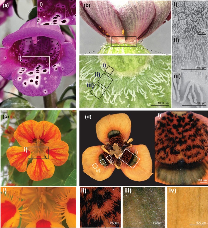
Variation in trichome‐like structures creates floral micropatterns. (a) Digitalis purpurea (Foxglove) with elongated trichomes on its corolla (i). (b) Three trichome domains in the floral nectary of Hibiscus trionum. (i) Nectar‐secreting trichomes closest to the base of the flower, (ii) single‐celled defensive trichomes in the middle region and (iii) enlarged glandular trichomes at the periphery of the nectary. (c) The petal of Tropaeolum majus (Nasturtium) with (i) serration‐like outgrowths. (d) The petal of Moraea tulbaghensis with different epidermal cell types in three discrete domains (i). (ii) Cilia‐like structures in the proximal region, (iii) iridescent conical cells in the middle region and (iv) elongated yellow cells at the distal region of the petal.
Compared with leaves, the mechanisms specifying trichome identity and distribution on petals are not well understood. Investigations in a few species identified HD‐ZIP and MYB transcription factors as key regulators (Wu et al., 2023; Zahid et al., 2023), but it is not clear whether distinct trichome types are regulated by distinct MYBs or whether a single MYB can produce different trichomes types when the timing or level of its expression, or the availability of its co‐regulators vary. Studies on petal trichomes suggest that the regulators involved differ from that of other organs: While subgroup 9 MYBs (MIXTA‐like) regulate hair fate on the corolla of several species (e.g. Tan et al., 2016; Chen & Yuan, 2024), leaf trichome development is controlled by MYB subgroup 15 members, at least in Arabidopsis. Specialization within subgroup 9 has occurred to regulate petal (GhMYB10) and seed trichomes (GhMYB25 and GhMYB25‐like) in cotton, suggesting that gene duplication and functionalization could be instrumental in the diversification of trichome type. Reciprocally, the tufted trichomes (unicellular elongated hairs) at the base of the cotton petal are regulated by GhMYB25 and GhMYB25‐like, and subsequent co‐option of the associated regulatory network in the seed epidermis could be a first step towards the evolution of cotton fibres (Tan et al., 2020).
Assemblages of petal trichomes form nectar guides in Mimulus, but what controls interspecific variation in this trait was not understood. Recent findings start to fill the gap, showing that GUIDELESS, a subgroup 9 MIXTA‐like MYB, underpins differences in trichome length between the nectar guides of two Mimulus species (Chen & Yuan, 2024). In the bumblebee‐pollinated M. lewisii, GUIDELESS promotes the production of long trichomes along the nectar guides. In the self‐pollinated M. parishii, a deleterious SNP in GUIDELESS yields shorter trichomes. However, the mechanism enabling GUIDELESS to regulate trichome length is still not understood. In addition, GUIDELESS is not the only locus regulating nectar guide trichome length between these species, suggesting that other unknown genetic factors have contributed to a switch in mating strategy from bumblebee‐mediated pollination to selfing.
Hook‐shaped trichomes on the surface of the petal‐like sepals of Aristolochia esperanzae are central to pollen removal and fruit set: shaved flowers capture < 1% of the pollinators caught by a trichome‐bearing flower and fail to produce fruits (Matallana‐Puerto et al., 2024). Trichomes distribution, orientation and morphology directly contribute to the efficiency of the trap. However, a closely related species, A. macrophylla, lacks such pollen‐trapping trichomes, suggesting that there could be other evolutionarily significance to their presence or absence (Suárez‐Baron et al., 2023). Elucidating the mechanisms governing patterning of these trichomes should contribute to our understanding of a structure that directly affects reproductive fitness: transcriptomic studies identified various MYBs (AfimGL2, AfimMYB106‐like) and members of the AP2/EREBP family (AfimRAV1‐like and AfimWIN1) as candidate to control trichome development on the perianth of A. fimbriata flowers (Suárez‐Baron et al., 2021). However, further work is necessary to functionally validate those genes and determine whether they could also regulate trichomes specification in petals and petaloid sepals of other species.
Finally, certain trichomes specialize in surprising ways: In cotton, trichomes on the outer epidermis of the corolla act as glue that holds adjacent petals together. These trichomes fulfil a mechanical role, ensuring correct bud shape and protecting reproductive organs from desiccation during development (Tan et al., 2016). The force required to separate petals from each other quantitatively relates to the extent of entanglement between stellate trichomes on adjacent petals. Correct patterning of cell fate is essential to achieve this ‘velcro effect’ as trichomes must reside on the part of the abaxial epidermis in direct contact with neighbouring petals, but here too, mechanisms controlling patterning are unknown.
Due to their diversity in both type and location, petal hairs provide a great opportunity to start exploring the role physical forces play in the evolution of flower form – an area currently understudied.
VI. Patterning cell behaviour supports the evolution of novel petal‐associated features
Petals in many species have highly elaborate shapes and appendages (Fig. 5c,d) that can be extraordinarily diverse (Endress & Matthews, 2006). These structures are excellent systems to explore general patterns of flowering plant diversification and understand evolution innovates, yet how they emerge is mostly unknown. How morphological novelties originate is an important question in evolutionary biology (Wagner, 2015), as evolutionary innovation led to novel functional capacities or new body parts. Controlling cell behaviour spatially across the corolla is essential for both and below we consider two specific cases.
Nectar producing glands, or nectaries, have a direct impact on pollinator attraction and contribute to speciation and evolution (Katzer et al., 2019; Parachnowitsch et al., 2019). Nectaries have evolved numerous times independently across angiosperms and are extremely diverse in respect to their location, structures and secretion mechanisms (Heil, 2011; Liao et al., 2025). The production of nectar relies on the development of specialized cell types, allowing us to explore evolutionary innovation at the cellular scale. In some species, trichome assemblage can form the basis of nectaries. For example, the nectaries of H. trionum and other Malvaceae are made of two concentric rings of hairs adjacent to the abaxial petal surface (Fig. 5d; Hu et al., 2020). Glandular trichomes closest to the petal base produce nectar while the outer whorl of uniseriate trichomes might play a defensive role (Fig. 5d). Elucidating the processes mediating the specification of two distinct hair types next to each other to generate a multifunctional structure represents a promising venue to understand how evolution generates a novel capacity (i.e. the ability to produce nectar). It will also be interesting to determine whether the mechanisms that mediate variation in nectary size and nectar volume produced by a flower is associated with pollination syndrome (Katzer et al., 2019).
In some species, nectar is produced at the bottom of long tubular outgrowths that restrict its access to visitors with a tongue long enough to reach it. These structures, called nectar spurs, can be derived from multiple floral organs (petals and sepals) and have evolved multiple times independently across diverse species of angiosperms (Hodges, 1997; Li et al., 2024). Morphological variation in the petal‐derived nectar spurs in Aquilegia can impact pollination: Long nectar spurs in North American populations of Aquilegia are associated with a reduction in pigment and gain of hawkmoths pollinators (Whittall & Hodges, 2007). In Asia, Aquilegia have evolved shorter nectar spurs while shifting to primarily bumblebee and scarid fly pollinators (Tang et al., 2007). Switching to the specialist hawkmoth as primary pollinator promotes outcrossing over the tendency for self‐pollination by more generalist bumblebees, suggesting that trade‐offs for the gain or loss of spurs impact reproductive isolation and speciation (Brunet & Sweet, 2006).
Nectar spurs form when a group of cells in the emerging organ primordia experience enhanced cell division compared with the surrounding tissues (Fig. 6a). Subsequent spur elongation is driven through cell expansion or cell proliferation and different species use distinct strategies, and different genes, to achieve the final spur size. Cell length and anisotropy correlate with spur length in the Aquilegia genus (Puzey et al., 2012), but in Linaria species, cell size appears similar between species, and variation in spur dimension is due to differences in cell number (Cullen et al., 2018; Fig. 6b). This indicates that either process can be modulated to control spur dimensions. Remarkably, constitutive overexpression of a KNOX transcription factor associated with spur growth in snapdragon and tobacco, two spurless species, is sufficient to produce spur‐like appendages (Fig. 6c; Box et al., 2011), indicating that while these species lack the ability to grow spurs, they possess the programme allowing a group of cells to ‘break free’ from the rest of the petal. This suggests petal spur origination required two key events: the ability to specify the ‘spur spot’, to allow a group of cells to behave independently (individuation), followed by the independent recruitment of diverse ‘growth modules’ (genes controlling cell proliferation and/or expansion in the spur spot) along distinct lineages (Fig. 6a).
Fig. 6.

Individuation and change in growth at the cellular scale can support the evolution of novelty and biodiversity. (a) Nectar spur specification allows a subdomain of the petal primordia to become differentiated (individuation) and a spur emerges as cell proliferation is promoted in this ‘spur domain’. Depending on the genus, spur elongation is mostly driven through cell proliferation or cell expansion. (b) Interspecific variation in spur length is due to changes in cell expansion (differences in cell length/anisotropy) in Aquilegia (Puzey et al., 2012) and variation in cell proliferation (no difference in cell length but difference in number of cells) in Linaria (Cullen et al., 2018). Red arrows depict the average cell length in each case (c) Spur formation (black arrow) can be induced in spurless species, such as snapdragon (another species of Lamiales) or tobacco (Solanales), when a KNOX transcription factor promoting cell growth is constitutively overexpressed (Box et al., 2011).
While KNOX genes promote cell proliferation and spur elongation in Lamiales, other species use distinct regulators to control spur growth. Mapping spur length in Aquilegia found that POPOVICH (POP), a zinc finger domain transcription factor, promotes cell proliferation during the early stages of spur development and loss‐of‐function mutations in POP accounts for loss of spurs in Aquilegia ecalcarata, suggesting that this locus is also an important contributor to petal spur evolution (Min et al., 2019; Ballerini et al., 2020). Spur elaboration in Aquilegia also depends on local repression of cell division by TEOSINTE‐BRANCHED‐like transcription factors (TCP4) and cell elongation likely driven by complex hormone signalling, including cytokinin, auxin, gibberellin and brassinosteroids (Yant et al., 2015; Conway et al., 2021). These pathways could also be involved in the evolution of nectar spurs in other species, as TCP‐like genes and hormonal responses have been proposed to regulate nectar spur length in Linaria and Antirrhinum, and the sepal/petal‐derived spurs of Tropaeolum (Cullen et al., 2023; Martínez‐Salazar et al., 2023).
However, the nature and origination of the signalling events that specify the spur spot in the first place are still poorly understood. To which extent those individuation processes merge with the prepatterning events that divide the petal surface allowing neighbouring cells to acquire distinct fates (see Section III and Fig. 4g) remains to be investigated. Resolving the mechanisms that control cell behaviour (growth and differentiation) across the petal with such spatial precision to produce autonomous subdomains would not only allow us to understand spur initiation but also shed light on events that enable evolution to innovate and create new body parts.
VII. Drivers of natural selection on petal patterns and their contribution to speciation
Because change in patterns across the petal surface directly impacts plant reproductive success, variation in the processes that govern cell behaviour (growth and differentiation) and the emergence of these patterns constitutes a useful system for understanding the evolution of complex traits. However, there are limited studies that examine the level of selection on these traits across the genome. Here, we summarize what is known about the evolution of petal pigmentation, and how it can help us understand inter‐ and intraspecific adaptation and evolution.
1. Evolution of novel genetic material and selection of petal traits
For selection to act upon phenotypic variation, genetic variation must first emerge. Gene duplication and neofunctionalization are two main drivers of genome evolution (Clark, 2023). Gene duplication allows for the creation of new genetic material and yield paralogs that can evolve new functions. Indeed several transcription factor families have undergone extensive gene duplication and sequence divergence across the angiosperms (Irish, 2003; Zhang et al., 2023). Its impact on the diversification of petal patterns has been discussed throughout this review, mostly through cases from the MYB transcription factor family. Transposons are also a driver of evolution through the creation of genetic diversity across eukaryotes, and unsurprisingly, they also account for variation in petal pigmentation. Gene disruptions via transposons account for anthocyanin modification in Petunia (Nakajima et al., 2005) and rose (Li et al., 2022), white flower pigmentation in Chinese kale (Zhang et al., 2019), and variegated pigmentation patterns in varieties of Japanese morning glory (Iida et al., 2004).
Genome structure can facilitate reproductive isolation of closely related species by linking several genes that control species‐specific traits, insuring morphological characteristics segregate together (Gutiérrez‐Valencia et al., 2021). These speciation ‘super loci’ have been found in Petunia, Mimulus and Anthurium, and contain several genes that regulate key differences between species. For example, in Petunia, multiple traits associated with flower patterning and pollinator syndrome, including UV absorption, scent production and pistil length, are tightly linked, insuring they are inherited together (Hermann et al., 2013; Amrad et al., 2016). More recently, this locus was found to contain a gene that causes hybrid necrosis in plants that carry incompatible alleles, causing hybrids between Petunia species to be stunted and further promoting their separation in nature (Li et al., 2023). A similar phenomenon was observed in Mimulus. The YUP siRNA locus in M. parishii and M. cardinalis mentioned above is linked to two other MYB transcription factors genes SISTER OF LIGHT AREAS1 (SOLAR) and PETAL LOBE ANTHOCYANIN (PELAN), both of which control aspects of petal pigmentation that influence pollinator preferences (Liang et al., 2023). Such ‘super loci’ provide an interesting mechanism for how closely related species can prevent cross pollination and hybridization in nature. Together, these findings help us understand how petal pigmentation patterns can contribute to reproductive isolation in natural populations and act as drivers of speciation events.
2. Evolution in situ: the importance of examining petal patterns in natural populations
To date, little has been done to understand how the genes regulating petal patterning behave under selection in natural populations. However, a handful of field studies underscore the contribution of patterned cell behaviour to evolution and speciation. For example, the genetic loci and networks regulating pigmentation in Antirrhinum majus are well understood. This basic knowledge has facilitated field studies to examine gene flow and natural selection in wild populations (Field et al., 2025). A recent study surveyed a natural hybrid zone that contains two subspecies of A. majus with distinct petal patterns. Pooled whole‐genome sequencing revealed that selective sweeps on two tightly linked MYB transcriptions factors that regulate pigment, EL and ROS, have prevented gene flow between distinct populations. These loci allow for discrete petal pigment phenotypes to be maintained and provide a potential mechanism for reproductive isolation and speciation in these populations (Tavares et al., 2018).
Examining nectar spur evolution in Aquilegia coerulea has revealed that gain or loss of nectar spurs can happen in a short time frame because of trade‐offs between pollinator preference and herbivory. In Aquilegia, the presence of long nectar spurs is an adaptation to pollination by hawkmoths, as their probiscis can reach nectar reward at the end of the spur (Whittall & Hodges, 2007). Analysis of linkage disequilibrium in natural populations of A. coerulea in North America uncovered a QTL containing loss‐of function mutations in an APETALA3‐3‐like gene that causes petal‐like nectar spurs to be replaced with sepals (Cabin et al., 2022). This locus was under selection in areas with increased herbivory, suggesting a fitness advantage for these homeotic mutants. However, it also caused a shift in pollinator preference from hawkmoth to bumblebees, resulting in a decrease in outcrossing rates in populations containing the spurless morphology. This suggests that this locus is contributing to reproductive isolation in these Aquilegia populations, and it will be interesting to see whether these two floral morphologies lead to a speciation event in the future (Cabin et al., 2022). Together, these examples show how complex petal traits are evolving in response to changing environments, and highlights the importance of using foundational knowledge of developmental programmes to understand how they are altered in naturally evolving populations in real time.
VIII. Conclusion and insights
Petals display various cellular arrangements that directly impact plant fitness, making them powerful systems to decipher how the interplay between evolutionary and developmental processes shapes the history of flowering plants. While significant progress has been made in understanding the genetic mechanisms and adaptive benefits of some of these cellular features like pigmentation patterns, other traits – such as cell shape variation, cuticle texture and the specification of various floral trichome types – require further characterization.
In addition, outstanding questions remain. For example, how such diverse cell types are specified within the same organ is far from understood. MYB transcription factors regulate tissue‐specific pigment production and, in some species, also influence cell shape or cuticle texture. However, how they regulate diverse traits across species and what controls their spatiotemporal expression remain elusive (Fig. 4). While recent studies in Mimulus and Antirrhinum hint at possible answers, such as the role of regulatory RNAs in controlling colour pattern production, whether this phenomenon applies across a broader range of species in not known. More importantly, upstream events that create subdomains within the petal primordia to allow neighbouring cells to embark on distinct growth and differentiation trajectories (Figs 4h, 6a) are still unknown, and solving this enigma constitutes an exciting challenge for the field.
Here, we argue that one way to answer some of these questions is by exploiting natural genetic and phenotypic variation across diverse species. The value of wild morphological variation has been highlighted in an increasing number of field studies and is certain to provide valuable insights into the vast diversity of flowering plants. Future research should focus on characterizing petal cell traits in situ, sequencing collections of novel ecotypes and developing robust transformation protocols for species well‐suited to investigate understudied petal features.
As we continue to unravel the genetic networks and key regulatory players of petal patterning, the integration of developmental genetics, genomics, high‐resolution imaging, mathematical modelling and ecological surveys will be crucial for advancing our understanding of petal patterning at the cellular level. We believe such interdisciplinary approaches are pivotal to move the field beyond descriptive studies to gain mechanistic insights on the complexity of patterning the microscale and its contribution to the evolution and diversification of flowering plants at the macroscale.
Competing interests
None declared.
Author contributions
ED and EM discussed ideas, prepared figures and wrote the manuscript.
Disclaimer
The New Phytologist Foundation remains neutral with regard to jurisdictional claims in maps and in any institutional affiliations.
Acknowledgements
We would like to thank Prof. Elena Kramer and Prof. Liam Dolan for the opportunity to write this review as well as three anonymous reviewers for their constructive and detailed comments on a previous version of this manuscript. We also thank Dr Raymond Wightman for assistance with the cryo‐electron microscopy, the Cambridge University Botanical Garden (CUBG) for access to their botanical collections, May Yeo for flower images and Dr Lucie Riglet for artificial flowers. Research in our laboratory related to the topic of this review is funded by the Gatsby Charitable Foundation (grant nos.: RG92362 and G117782) and the Isaac Newton/ Wellcome Fund (grant no.: RG89529).
References
- Abrahamczyk S, Humphreys AM, Trabert F, Droppelmann F, Gleichmann M, Krieger V, Linnartz M, Lozada‐Gobilard S, Rahelivololona ME, Schubert M et al. 2021. Evolution of brood‐site mimicry in Madagascan impatiens (Balsaminaceae). Perspectives in Plant Ecology, Evolution and Systematics 49: 125590. [Google Scholar]
- Airoldi CA, Lugo CA, Wightman R, Glover BJ, Robinson S. 2021. Mechanical buckling can pattern the light‐diffracting cuticle of Hibiscus trionum . Cell Reports 36: 109715. [DOI] [PMC free article] [PubMed] [Google Scholar]
- Alonso‐Blanco C, Andrade J, Becker C, Bemm F, Bergelson J, Borgwardt KM, Cao J, Chae E, Dezwaan TM, Ding W et al. 2016. 1,135 genomes reveal the global pattern of polymorphism in Arabidopsis thaliana . Cell 166: 481–491. [DOI] [PMC free article] [PubMed] [Google Scholar]
- Amrad A, Moser M, Mandel T, de Vries M, Schuurink RC, Freitas L, Kuhlemeier C. 2016. Gain and loss of floral scent production through changes in structural genes during pollinator‐mediated speciation. Current Biology 26: 3303–3312. [DOI] [PubMed] [Google Scholar]
- Antoniou Kourounioti RL, Band LR, Fozard JA, Hampstead A, Lovrics A, Moyroud E, Vignolini S, King JR, Jensen OE, Glover BJ. 2013. Buckling as an origin of ordered cuticular patterns in flower petals. Journal of the Royal Society Interface 10: 20120847. [DOI] [PMC free article] [PubMed] [Google Scholar]
- Baczyński J, Celep F, Spalik K, Claßen‐Bockhoff R. 2022. Flower‐like meristem conditions and spatial constraints shape architecture of floral pseudanthia in Apioideae. EvoDevo 13: 19. [DOI] [PMC free article] [PubMed] [Google Scholar]
- Bailes EJ, Pattrick JG, Glover BJ. 2018. An analysis of the energetic reward offered by field bean (Vicia faba) flowers: nectar, pollen, and operative force. Ecology and Evolution 8: 3161–3171. [DOI] [PMC free article] [PubMed] [Google Scholar]
- Ballerini ES, Min Y, Edwards MB, Kramer EM, Hodges SA. 2020. POPOVICH, encoding a C2H2 zinc‐finger transcription factor, plays a central role in the development of a key innovation, floral nectar spurs, in Aquilegia. Proceedings of the National Academy of Sciences, USA 117: 22552–22560. [DOI] [PMC free article] [PubMed] [Google Scholar]
- von Balthazar M, Lachmayer M, Hawranek A‐S, Kopper C, Schönenberger J, Chartier M. 2025. Pollination and reproductive systems in Columbines (Aquilegia, Ranunculaceae): review and Insights. International Journal of Plant Sciences 186: 38–52. [Google Scholar]
- Barrett SCH. 2010. Darwin's legacy: the forms, function and sexual diversity of flowers. Philosophical Transactions of the Royal Society, B: Biological Sciences 365: 351–368. [DOI] [PMC free article] [PubMed] [Google Scholar]
- Becker A, Yamada Y, Sato F. 2023. California poppy (Eschscholzia californica), the Papaveraceae golden girl model organism for evodevo and specialized metabolism. Frontiers in Plant Science 14: 1084358. [DOI] [PMC free article] [PubMed] [Google Scholar]
- Berry E, Choudhary AK, Geeta R. 2023. Why do some funneliform flowers have petal folds accompanied with hierarchical surface microstructure? Evolutionary Ecology 37: 385–399. [Google Scholar]
- Berry E, Geeta R. 2019. Variation in surface microstructure of the corolla with respect to pigment pattern in some Acanthaceae. Phytomorphology 69: 25–31. [Google Scholar]
- Bowman JL, Moyroud E. 2024. Reflections on the ABC model of flower development. Plant Cell 36: 1334–1357. [DOI] [PMC free article] [PubMed] [Google Scholar]
- Bowman JL, Smyth DR, Meyerowitz EM. 2012. The ABC model of flower development: then and now. Development 139: 4095–4098. [DOI] [PubMed] [Google Scholar]
- Box MS, Dodsworth S, Rudall PJ, Bateman RM, Glover BJ. 2011. Characterization of Linaria KNOX genes suggests a role in petal‐spur development. The Plant Journal 68: 703–714. [DOI] [PubMed] [Google Scholar]
- Bradley D, Boell L, Richardson D, Copsey L, Whibley A, Xu T, Zhang Y, Xue Y, Field D, Coen E. 2025. Shaping of developmental gradients through selection on multiple loci in Antirrhinum. bioRxiv . doi: 10.1101/2025.02.12.637738. [DOI]
- Bradley D, Xu P, Mohorianu I‐I, Whibley A, Field D, Tavares H, Couchman M, Copsey L, Carpenter R, Li M et al. 2017. Evolution of flower color pattern through selection on regulatory small RNAs. Science 358: 925–928. [DOI] [PubMed] [Google Scholar]
- Bradshaw HD, Schemske DW. 2003. Allele substitution at a flower colour locus produces a pollinator shift in monkeyflowers. Nature 426: 176–178. [DOI] [PubMed] [Google Scholar]
- Bräuer P, Neinhuis C, Voigt D. 2017. Attachment of honeybees and greenbottle flies to petal surfaces. Arthropod‐Plant Interactions 11: 171–192. [Google Scholar]
- Brockington SF, Alvarez‐Fernandez R, Landis JB, Alcorn K, Walker RH, Thomas MM, Hileman LC, Glover BJ. 2013. Evolutionary analysis of the MIXTA gene family highlights potential targets for the study of cellular differentiation. Molecular Biology and Evolution 30: 526–540. [DOI] [PubMed] [Google Scholar]
- Brunet J, Sweet HR. 2006. Impact of insect pollinator group and floral display size on outcrossing rate. Evolution 60: 234–246. [PubMed] [Google Scholar]
- Cabin Z, Derieg NJ, Garton A, Ngo T, Quezada A, Gasseholm C, Simon M, Hodges SA. 2022. Non‐pollinator selection for a floral homeotic mutant conferring loss of nectar reward in Aquilegia coerulea . Current Biology 32: 1332–1341. [DOI] [PubMed] [Google Scholar]
- Cavallini‐Speisser Q, Morel P, Monniaux M. 2021. Petal cellular identities. Frontiers in Plant Science 12: 4589. [DOI] [PMC free article] [PubMed] [Google Scholar]
- Chen H, Yuan Y‐W. 2024. Genetic basis of nectar guide trichome variation between bumblebee‐ and self‐pollinated monkeyflowers (Mimulus): role of the MIXTA‐like gene GUIDELESS. BMC Plant Biology 24: 62. [DOI] [PMC free article] [PubMed] [Google Scholar]
- Cheng J, Yao X, Li X, Yue L, Duan X, Li B, Fu X, Li S, Shan H, Yin X et al. 2023. Diversification of ranunculaceous petals in shape supports a generalized model for plant lateral organ morphogenesis and evolution. Science Advances 9: eadf8049. [DOI] [PMC free article] [PubMed] [Google Scholar]
- Chopy M, Cavallini‐Speisser Q, Chambrier P, Morel P, Just J, Hugouvieux V, Rodrigues Bento S, Zubieta C, Vandenbussche M, Monniaux M. 2023. Cell layer–specific expression of the homeotic MADS‐box transcription factor PhDEF contributes to modular petal morphogenesis in petunia. Plant Cell 36: 324–345. [DOI] [PMC free article] [PubMed] [Google Scholar]
- Christensen KI, Hansen HV. 1998. SEM‐studies of epidermal patterns of petals in the angiosperms. Opera Botanica 135: 4589. [Google Scholar]
- Clark JW. 2023. Genome evolution in plants and the origins of innovation. New Phytologist 240: 2204–2209. [DOI] [PubMed] [Google Scholar]
- Conway SJ, Walcher‐Chevillet CL, Salome Barbour K, Kramer EM. 2021. Brassinosteroids regulate petal spur length in Aquilegia by controlling cell elongation. Annals of Botany 128: 931–942. [DOI] [PMC free article] [PubMed] [Google Scholar]
- Costa VBS, Pimentel RMM, Chagas MGS, Alves GD, Castro CC. 2017. Petal micromorphology and its relationship to pollination. Plant Biology 19: 115–122. [DOI] [PubMed] [Google Scholar]
- Cullen E, Fernández‐Mazuecos M, Glover BJ. 2018. Evolution of nectar spur length in a clade of Linaria reflects changes in cell division rather than in cell expansion. Annals of Botany 122: 801–809. [DOI] [PMC free article] [PubMed] [Google Scholar]
- Cullen E, Wang Q, Glover BJ. 2023. How do you build a nectar spur? A transcriptomic comparison of nectar spur development in Linaria vulgaris and gibba development in Antirrhinum majus . Frontiers in Plant Science 14: 4589. [DOI] [PMC free article] [PubMed] [Google Scholar]
- Damerval C, Becker A. 2017. Genetics of flower development in Ranunculales – a new, basal eudicot model order for studying flower evolution. New Phytologist 216: 361–366. [DOI] [PubMed] [Google Scholar]
- Davies KM, Albert NW, Zhou Y, Schwinn KE. 2018. Functions of Flavonoid and Betalain Pigments in Abiotic Stress Tolerance in Plants. In: Annual Plant Reviews online. New York, NY, USA: John Wiley & Sons, Ltd, 21–62. [Google Scholar]
- Di Stilio VS, Martin C, Schulfer AF, Connelly CF. 2009. An ortholog of MIXTA‐like2 controls epidermal cell shape in flowers of Thalictrum . New Phytologist 183: 718–728. [DOI] [PubMed] [Google Scholar]
- Ding B, Patterson EL, Holalu SV, Li J, Johnson GA, Stanley LE, Greenlee AB, Peng F, Bradshaw HD, Blinov ML et al. 2020. Two MYB proteins in a self‐organizing activator‐inhibitor system produce spotted pigmentation patterns. Current Biology 30: 802–814. [DOI] [PMC free article] [PubMed] [Google Scholar]
- Duan A‐Q, Deng Y‐J, Liu H, Xu Z‐S, Xiong A‐S. 2024. An anthocyanin activation gene underlies the purple central flower pigmentation in wild carrot. Plant Physiology 196: 1147–1162. [DOI] [PubMed] [Google Scholar]
- Durán‐Castillo M, Hudson A, Wilson Y, Field DL, Twyford AD. 2021. A phylogeny of Antirrhinum reveals parallel evolution of alpine morphology. New Phytologist 233: 1426–1439. [DOI] [PubMed] [Google Scholar]
- Edwards MB, Ballerini ES, Kramer EM. 2022. Complex developmental and transcriptional dynamics underlie pollinator‐driven evolutionary transitions in nectar spur morphology in Aquilegia (columbine). American Journal of Botany 109: 1360–1381. [DOI] [PubMed] [Google Scholar]
- Ellis AG, Johnson SD. 2009. The evolution of floral variation without pollinator shifts in Gorteria diffusa (Asteraceae). American Journal of Botany 96: 793–801. [DOI] [PubMed] [Google Scholar]
- Endress PK. 2001. Origins of flower morphology. The Journal of Experimental Zoology 291: 105–115. [DOI] [PubMed] [Google Scholar]
- Endress PK, Matthews ML. 2006. Elaborate petals and staminodes in eudicots: diversity, function, and evolution. Organisms Diversity & Evolution 6: 257–293. [Google Scholar]
- Fattorini R, Khojayori FN, Mellers G, Moyroud E, Herrero E, Kellenberger RT, Walker R, Wang Q, Hill L, Glover BJ. 2024. Complex petal spot formation in the beetle daisy (Gorteria diffusa) relies on spot‐specific accumulation of malonylated anthocyanin regulated by paralogous GdMYBSG6 transcription factors. New Phytologist 243: 240–257. [DOI] [PubMed] [Google Scholar]
- Field DL, Stankowski S, Reiter T, Polechova J, Bradley D, Richardson D, Pal A, Shipilina D, Boell L, Pickup M et al. 2025. Genome‐wide cline analysis identifies new locus contributing to a barrier to gene flow across an Antirrhinum hybrid zone. bioRxiv . doi: 10.1101/2025.02.17.638607. [DOI]
- Free JB. 1970. Effect of flower shapes and nectar guides on the behaviour of foraging honeybees. Behaviour 37: 269–285. [Google Scholar]
- Fujii T, Matsuda S, Tejedor ML, Esaki T, Sakane I, Mizuno H, Tsuyama N, Masujima T. 2015. Direct metabolomics for plant cells by live single‐cell mass spectrometry. Nature Protocols 10: 1445–1456. [DOI] [PubMed] [Google Scholar]
- Galipot P, Damerval C, Jabbour F. 2021. The seven ways eukaryotes produce repeated colour motifs on external tissues. Biological Reviews 96: 1676–1693. [DOI] [PubMed] [Google Scholar]
- Gao Y, Li J, He J, Yu Y, Qian Z, Geng Z, Yang L, Zhang Y, Ke Y, Lin Q et al. 2024. BLADE‐ON‐PETIOLE interacts with CYCLOIDEA to fine‐tune CYCLOIDEA ‐mediated flower symmetry in monkeyflowers (Mimulus). Science Advances 10: eado4571. [DOI] [PMC free article] [PubMed] [Google Scholar]
- Gaskett AC. 2011. Orchid pollination by sexual deception: pollinator perspectives. Biological Reviews 86: 33–75. [DOI] [PubMed] [Google Scholar]
- Ghissing U, Mitra A. 2022. Biology of floral scent volatiles in ornamental plants. In: Datta SK, Gupta YC, eds. Floriculture and ornamental plants. Singapore city, Singapore: Springer Nature, 777–817. [Google Scholar]
- Giorio C, Moyroud E, Glover BJ, Skelton PC, Kalberer M. 2015. Direct surface analysis coupled to high‐resolution mass spectrometry reveals heterogeneous composition of the cuticle of Hibiscus trionum petals. Analytical Chemistry 87: 9900–9907. [DOI] [PubMed] [Google Scholar]
- Gorb EV, Gorb SN. 2023. Petals reduce attachment of insect pollinators: a case study of the plant Dahlia pinnata and the fly Eristalis tenax . Insects 14: 285. [DOI] [PMC free article] [PubMed] [Google Scholar]
- Green AA, Kennaway JR, Hanna AI, Bangham JA, Coen E. 2010. Genetic control of organ shape and tissue polarity. PLoS Biology 8: e1000537. [DOI] [PMC free article] [PubMed] [Google Scholar]
- Guo Y, Chen X, Li J, Wang Q, Zhang S, Liu N, Zhang Y, Zhang T. 2024. Single‐cell RNA sequencing reveals a high‐resolution cell atlas of petals in Prunus mume at different flowering development stages. Horticulture Research 11: uhae189. [DOI] [PMC free article] [PubMed] [Google Scholar]
- Gutiérrez‐Valencia J, Hughes PW, Berdan EL, Slotte T. 2021. The genomic architecture and evolutionary fates of supergenes. Genome Biology and Evolution 13: evab057. [DOI] [PMC free article] [PubMed] [Google Scholar]
- Hatlestad GJ, Akhavan NA, Sunnadeniya RM, Elam L, Cargile S, Hembd A, Gonzalez A, McGrath JM, Lloyd AM. 2015. The beet Y locus encodes an anthocyanin MYB‐like protein that activates the betalain red pigment pathway. Nature Genetics 47: 92–96. [DOI] [PubMed] [Google Scholar]
- Heil M. 2011. Nectar: generation, regulation and ecological functions. Trends in Plant Science 16: 191–200. [DOI] [PubMed] [Google Scholar]
- Hermann K, Klahre U, Moser M, Sheehan H, Mandel T, Kuhlemeier C. 2013. Tight genetic linkage of prezygotic barrier loci creates a multifunctional speciation island in petunia. Current Biology 23: 873–877. [DOI] [PubMed] [Google Scholar]
- Hodges SA. 1997. Floral nectar spurs and diversification. International Journal of Plant Sciences 158: S81–S88. [Google Scholar]
- Hsu H‐F, Chen W‐H, Shen Y‐H, Hsu W‐H, Mao W‐T, Yang C‐H. 2021. Multifunctional evolution of B and AGL6 MADS box genes in orchids. Nature Communications 12: 902. [DOI] [PMC free article] [PubMed] [Google Scholar]
- Hsu H‐F, Hsu W‐H, Lee Y‐I, Mao W‐T, Yang J‐Y, Li J‐Y, Yang C‐H. 2015. Model for perianth formation in orchids. Nature Plants 1: 1–8. [Google Scholar]
- Hu W, Qin W, Jin Y, Wang P, Yan Q, Li F, Yang Z. 2020. Genetic and evolution analysis of extrafloral nectary in cotton. Plant Biotechnology Journal 18: 2081–2095. [DOI] [PMC free article] [PubMed] [Google Scholar]
- Iida S, Morita Y, Choi J‐D, Park K‐I, Hoshino A. 2004. Genetics and epigenetics in flower pigmentation associated with transposable elements in morning glories. Advances in Biophysics 38: 141–159. [PubMed] [Google Scholar]
- Irish VF. 2003. The evolution of floral homeotic gene function. BioEssays 25: 637–646. [DOI] [PubMed] [Google Scholar]
- Johnson SD, Midgley JJ. 1997. Fly pollination of Gorteria diffusa (Asteraceae), and a possible mimetic function for dark spots on the capitulum. American Journal of Botany 84: 429–436. [PubMed] [Google Scholar]
- Johnson SD, Schiestl FP. 2016. Floral mimicry. Oxford: Oxford University Press. [Google Scholar]
- Kang M, Choi Y, Kim H, Kim S‐G. 2022. Single‐cell RNA‐sequencing of Nicotiana attenuata corolla cells reveals the biosynthetic pathway of a floral scent. New Phytologist 234: 527–544. [DOI] [PMC free article] [PubMed] [Google Scholar]
- Katzer AM, Wessinger CA, Hileman LC. 2019. Nectary size is a pollination syndrome trait in Penstemon. New Phytologist 223: 377–384. [DOI] [PMC free article] [PubMed] [Google Scholar]
- Kay QON, Daoud HS, Stirton CH. 1981. Pigment distribution, light reflection and cell structure in petals. Botanical Journal of the Linnean Society 83: 57–83. [Google Scholar]
- Kellenberger RT, Ponraj U, Delahaie B, Fattorini R, Balk J, Lopez‐Gomollon S, Müller KH, Ellis AG, Glover BJ. 2023. Multiple gene co‐options underlie the rapid evolution of sexually deceptive flowers in Gorteria diffusa . Current Biology 33: 1502–1512.e8. [DOI] [PubMed] [Google Scholar]
- Kellogg EA. 2015. Brachypodium distachyonas a Genetic Model System. Annual Review of Genetics 49: 1–20. [DOI] [PubMed] [Google Scholar]
- Khojayori FN, Ponraj U, Buch K, Zhao Y, Herrera‐Ubaldo H, Glover BJ. 2024. Evolution and development of complex floral displays. Development 151: dev203027. [DOI] [PMC free article] [PubMed] [Google Scholar]
- Koch K, Bohn HF, Barthlott W. 2009. Hierarchically sculptured plant surfaces and Superhydrophobicity. Langmuir 25: 14116–14120. [DOI] [PubMed] [Google Scholar]
- Kolosova N, Sherman D, Karlson D, Dudareva N. 2001. Cellular and subcellular localization of S‐adenosyl‐l‐methionine: benzoic Acid Carboxyl methyltransferase, the enzyme responsible for biosynthesis of the volatile ester methylbenzoate in snapdragon flowers. Plant Physiology 126: 956–964. [DOI] [PMC free article] [PubMed] [Google Scholar]
- Koski MH, Ashman T‐L. 2015. Floral pigmentation patterns provide an example of Gloger's rule in plants. Nature Plants 1: 1–5. [DOI] [PubMed] [Google Scholar]
- Koski MH, MacQueen D, Ashman T‐L. 2020. Floral pigmentation has responded rapidly to global change in ozone and temperature. Current Biology 30: 4425–4431. [DOI] [PubMed] [Google Scholar]
- Kraaij M, van der Kooi CJ. 2020. Surprising absence of association between flower surface microstructure and pollination system. Plant Biology 22: 177–183. [DOI] [PMC free article] [PubMed] [Google Scholar]
- Kramer EM. 2009. Aquilegia: a new model for plant development, ecology, and evolution. Annual Review of Plant Biology 60: 261–277. [DOI] [PubMed] [Google Scholar]
- Kramer EM, Hodges SA. 2010. Aquilegia as a model system for the evolution and ecology of petals. Philosophical Transactions of the Royal Society, B: Biological Sciences 365: 477–490. [DOI] [PMC free article] [PubMed] [Google Scholar]
- Kunst L, Samuels L. 2009. Plant cuticles shine: advances in wax biosynthesis and export. Current Opinion in Plant Biology 12: 721–727. [DOI] [PubMed] [Google Scholar]
- Lam TH, Ho YB, Chen S. 1980. Observations on ultraviolet‐fluorescent trichomes on the tepals of narcissus flowers. Plant Science Letters 18: 115–120. [Google Scholar]
- León‐Osper M, Narbona E. 2022. Unravelling the mystery of red flowers in the Mediterranean Basin: how to be conspicuous in a place dominated by hymenopteran pollinators. Functional Ecology 36: 2774–2790. [Google Scholar]
- Li C, Binaghi M, Pichon V, Cannarozzi G, Brandão de Freitas L, Hanemian M, Kuhlemeier C. 2023. Tight genetic linkage of genes causing hybrid necrosis and pollinator isolation between young species. Nature Plants 9: 420–432. [DOI] [PMC free article] [PubMed] [Google Scholar]
- Li M, Zhang H, Yang Y, Wang H, Xue Z, Fan Y, Sun P, Zhang H, Zhang X, Jin W. 2022. Rosa1, a transposable element‐like insertion, produces red petal coloration in rose through altering RcMYB114 transcription. Frontiers in Plant Science 13: 857684. [DOI] [PMC free article] [PubMed] [Google Scholar]
- Li S, Fan J, Xue C, Shan H, Kong H. 2024. Spur development and evolution: an update. Current Opinion in Plant Biology 81: 102573. [DOI] [PubMed] [Google Scholar]
- Liang M, Chen W, LaFountain AM, Liu Y, Peng F, Xia R, Bradshaw HD, Yuan Y‐W. 2023. Taxon‐specific, phased siRNAs underlie a speciation locus in monkeyflowers. Science 379: 576–582. [DOI] [PMC free article] [PubMed] [Google Scholar]
- Liang M, Ringham L, Ye C, Yan X, Schaumburger N, Cieslak M, Blinov M, Prusinkiewicz P, Yuan Y‐W. 2025. From spots to stripes: evolution of pigmentation patterns in monkeyflowers via modulation of a reaction‐diffusion system and its prepatterns. bioRxiv . doi: 10.1101/2025.01.10.632501. [DOI]
- Liao H, Fu X, Zhao H, Cheng J, Zhang R, Yao X, Duan X, Shan H, Kong H. 2020. The morphology, molecular development and ecological function of pseudonectaries on Nigella damascena (Ranunculaceae) petals. Nature Communications 11: 1777. [DOI] [PMC free article] [PubMed] [Google Scholar]
- Liao IT, Gong Y, Kramer EM, Nikolov LA. 2025. The developmental basis of floral nectary diversity and evolution. New Phytologist 246: 2462–2477. [DOI] [PMC free article] [PubMed] [Google Scholar]
- Liao P, Ray S, Boachon B, Lynch JH, Deshpande A, McAdam S, Morgan JA, Dudareva N. 2021. Cuticle thickness affects dynamics of volatile emission from petunia flowers. Nature Chemical Biology 17: 138–145. [DOI] [PubMed] [Google Scholar]
- Lin R‐C, Rausher MD. 2021a. R2R3‐MYB genes control petal pigmentation patterning in Clarkia gracilis ssp. sonomensis (Onagraceae). New Phytologist 229: 1147–1162. [DOI] [PubMed] [Google Scholar]
- Lin R‐C, Rausher MD. 2021b. Ancient gene duplications, rather than polyploidization, facilitate diversification of petal pigmentation patterns in Clarkia gracilis (Onagraceae). Molecular Biology and Evolution 38: 5528–5538. [DOI] [PMC free article] [PubMed] [Google Scholar]
- Litt A, Kramer EM. 2010. The ABC model and the diversification of floral organ identity. Seminars in Cell & Developmental Biology 21: 129–137. [DOI] [PubMed] [Google Scholar]
- Lloyd A, Brockman A, Aguirre L, Campbell A, Bean A, Cantero A, Gonzalez A. 2017. Advances in the MYB–bHLH–WD Repeat (MBW) pigment regulatory model: addition of a WRKY factor and co‐option of an anthocyanin MYB for Betalain regulation. Plant & Cell Physiology 58: 1431–1441. [DOI] [PMC free article] [PubMed] [Google Scholar]
- Lu H‐C, Lam S‐H, Zhang D, Hsiao Y‐Y, Li B‐J, Niu S‐C, Li C‐Y, Lan S, Tsai W‐C, Liu Z‐J. 2022. R2R3‐MYB genes coordinate conical cell development and cuticular wax biosynthesis in Phalaenopsis aphrodite . Plant Physiology 188: 318–331. [DOI] [PMC free article] [PubMed] [Google Scholar]
- Lugo CA, Airoldi C, Chen C, Crosby AJ, Glover BJ. 2023. Morphoelastic modelling of pattern development in the petal epidermal cell cuticle. Journal of the Royal Society Interface 20: 20230001. [DOI] [PMC free article] [PubMed] [Google Scholar]
- Lunau K, De Camargo MGG, Brito VLG. 2024. Pollen, anther, stamen, and androecium mimicry. Plant Biology 26: 349–368. [DOI] [PubMed] [Google Scholar]
- Ma X, Shi J, Bänziger H, Sun Y, Guo Y, Liu Z, Johnson SD, Luo Y. 2016. The functional significance of complex floral colour pattern in a food‐deceptive orchid. Functional Ecology 30: 721–732. [Google Scholar]
- Mao W‐T, Hsu W‐H, Song J‐L, Yang C‐H. 2024. The HD‐ZIP II gene PaHAT14 increases cuticle deposition by downregulating ERF gene PaERF105 in Phalaenopsis . Plant and Cell Physiology 65: 1751–1768. [DOI] [PubMed] [Google Scholar]
- Marinho CR, Souza CD, Barros TC, Teixeira SP. 2014. Scent glands in legume flowers. Plant Biology 16: 215–226. [DOI] [PubMed] [Google Scholar]
- Martínez‐Harms J, Hadar R, Márquez N, Menzel R, Shmida A, Stavenga DG, Vorobyev M. 2020. enhanced uv‐reflection facilitated a shift in the pollination system of the red poppy, Papaver rhoeas (Papaveraceae). Plants 9: 927. [DOI] [PMC free article] [PubMed] [Google Scholar]
- Martínez‐Salazar S, Kramer EM, González F, Pabón‐Mora N. 2023. Spatio‐temporal expression of candidate genes for nectar spur development in Tropaeolum (Tropaeolaceae: Brassicales). Annals of Botany 132: 1205–1218. [DOI] [PMC free article] [PubMed] [Google Scholar]
- Matallana‐Puerto CA, Brito VLG, Kuster VC, Oliveira PE, Cardoso JCF. 2024. Sex, flies and flower trap: trapping trichomes and their function in pollination. Functional Ecology 38: 2261–2270. [Google Scholar]
- Meinhardt H. 2012. Turing's theory of morphogenesis of 1952 and the subsequent discovery of the crucial role of local self‐enhancement and long‐range inhibition. Interface Focus 2: 407–416. [DOI] [PMC free article] [PubMed] [Google Scholar]
- de Melo MC, Taucce PPG, Borba EL. 2011. Reproductive biology and isolation mechanisms in rupicolous species of the Acianthera prolifera complex (Orchidaceae) occurring in southeastern Brazil. Plant Systematics and Evolution 293: 161–176. [Google Scholar]
- Min Y, Bunn JI, Kramer EM. 2019. Homologs of the STYLISH gene family control nectary development in Aquilegia. New Phytologist 221: 1090–1100. [DOI] [PubMed] [Google Scholar]
- Misra BB, Assmann SM, Chen S. 2014. Plant single‐cell and single‐cell‐type metabolomics. Trends in Plant Science 19: 637–646. [DOI] [PubMed] [Google Scholar]
- Mizzotti C, Galliani BM, Masiero S. 2014. The backstage of the ABC model: the Antirrhinum majus contribution. Plant Biosystems – An International Journal Dealing with All Aspects of Plant Biology 148: 176–186. [Google Scholar]
- Morita Y, Ishiguro K, Tanaka Y, Iida S, Hoshino A. 2015. Spontaneous mutations of the UDP‐glucose: flavonoid 3‐O‐glucosyltransferase gene confers pale‐ and dull‐colored flowers in the Japanese and common morning glories. Planta 242: 575–587. [DOI] [PubMed] [Google Scholar]
- Moyers BT, Owens GL, Baute GJ, Rieseberg LH. 2017. The genetic architecture of UV floral patterning in sunflower. Annals of Botany 120: 39–50. [DOI] [PMC free article] [PubMed] [Google Scholar]
- Moyroud E, Airoldi CA, Ferria J, Giorio C, Steimer SS, Rudall PJ, Prychid CJ, Halliwell S, Walker JF, Robinson S et al. 2022. Cuticle chemistry drives the development of diffraction gratings on the surface of Hibiscus trionum petals. Current Biology 32: 5323–5334. [DOI] [PubMed] [Google Scholar]
- Moyroud E, Glover BJ. 2017. The evolution of diverse floral morphologies. Current Biology 27: R941–R951. [DOI] [PubMed] [Google Scholar]
- Moyroud E, Wenzel T, Middleton R, Rudall PJ, Banks H, Reed A, Mellers G, Killoran P, Westwood MM, Steiner U et al. 2017. Disorder in convergent floral nanostructures enhances signalling to bees. Nature 550: 469–474. [DOI] [PubMed] [Google Scholar]
- Muravnik LE, Kostina OV, Zaporozhets NL. 2022. Structure and functions of the glandular trichomes in three Arnica species (Asteraceae), depending on their location on leaves and flowers. Flora 290: 152047. [Google Scholar]
- Nakajima T, Matsubara K, Kodama H, Kokubun H, Watanabe H, Ando T. 2005. Insertion and excision of a transposable element governs the red floral phenotype in commercial petunias. Theoretical and Applied Genetics 110: 1038–1043. [DOI] [PubMed] [Google Scholar]
- Neumann M, Xu X, Smaczniak C, Schumacher J, Yan W, Blüthgen N, Greb T, Jönsson H, Traas J, Kaufmann K et al. 2022. A 3D gene expression atlas of the floral meristem based on spatial reconstruction of single nucleus RNA sequencing data. Nature Communications 13: 2838. [DOI] [PMC free article] [PubMed] [Google Scholar]
- Noda K, Glover BJ, Linstead P, Martin C. 1994. Flower colour intensity depends on specialized cell shape controlled by a Myb‐related transcription factor. Nature 369: 661–664. [DOI] [PubMed] [Google Scholar]
- Ojeda DI, Jaén‐Molina R, Santos‐Guerra A, Caujape‐Castells J, Cronk Q. 2017. Temporal, but not spatial, changes in expression patterns of petal identity genes are associated with loss of papillate conical cells and the shift to bird pollination in Macaronesian lotus (Leguminosae). Plant Biology 19: 420–427. [DOI] [PubMed] [Google Scholar]
- Ojeda DI, Valido A, Fernández de Castro AG, Ortega‐Olivencia A, Fuertes‐Aguilar J, Carvalho JA, Santos‐Guerra A. 2016. Pollinator shifts drive petal epidermal evolution on the Macaronesian Islands bird‐flowered species. Biology Letters 12: 20160022. [DOI] [PMC free article] [PubMed] [Google Scholar]
- Ojeda I, Francisco‐Ortega J, Cronk QCB. 2009. Evolution of petal epidermal micromorphology in Leguminosae and its use as a marker of petal identity. Annals of Botany 104: 1099–1110. [DOI] [PMC free article] [PubMed] [Google Scholar]
- Oshima Y, Shikata M, Koyama T, Ohtsubo N, Mitsuda N, Ohme‐Takagi M. 2013. MIXTA‐Like Transcription Factors and WAX INDUCER1/SHINE1 coordinately regulate cuticle development in Arabidopsis and Torenia fournieri . Plant Cell 25: 1609–1624. [DOI] [PMC free article] [PubMed] [Google Scholar]
- Oud S, Żurowski MM, Jagt PL, van der Jagt S, Yeo MTS, Walker JF, Moyroud E, Vroomans RMA. 2025. Recurrent emergence of boundary cell types during evolution of floral bullseye patterns. bioRxiv . doi: 10.1101/2025.05.03.651954. [DOI]
- Pansarin ER, Maciel AA. 2017. Evolution of pollination systems involving edible trichomes in orchids. AoB Plants 9: plx033. [Google Scholar]
- Parachnowitsch AL, Manson JS, Sletvold N. 2019. Evolutionary ecology of nectar. Annals of Botany 123: 247–261. [DOI] [PMC free article] [PubMed] [Google Scholar]
- Patterson E, MacGregor DR, Heeney MM, Gallagher J, O'Connor D, Nuesslein B, Bartlett ME. 2024. Developmental constraint underlies the replicated evolution of grass awns. New Phytologist 245: 835–848. [DOI] [PMC free article] [PubMed] [Google Scholar]
- Peach K, Liu JW, Mazer SJ. 2020. Climate predicts UV floral pattern size, anthocyanin concentration, and pollen performance in Clarkia unguiculata . Frontiers in Plant Science 11: 847. [DOI] [PMC free article] [PubMed] [Google Scholar]
- Peng F, Sun X, van Vloten C, Correll J, Langdon M, Ngochanthra W, Johnson K, Amador Kane S. 2024. Hybrid mimulus flowers attract a new pollinator. New Phytologist 242: 1324–1332. [DOI] [PubMed] [Google Scholar]
- Poethig RS, Fouracre J. 2024. Temporal regulation of vegetative phase change in plants. Developmental Cell 59: 4–19. [DOI] [PMC free article] [PubMed] [Google Scholar]
- Pollack AJ, Gong X, Pollack JR. 2019. A common phytoene synthase mutation underlies white petal varieties of the California poppy. Scientific Reports 9: 11615. [DOI] [PMC free article] [PubMed] [Google Scholar]
- Pramanik D, Dorst N, Meesters N, Spaans M, Smets E, Welten M, Gravendeel B. 2020. Evolution and development of three highly specialized floral structures of bee‐pollinated Phalaenopsis species. EvoDevo 11: 16. [DOI] [PMC free article] [PubMed] [Google Scholar]
- Prüm B, Seidel R, Bohn HF, Speck T. 2011. Plant surfaces with cuticular folds are slippery for beetles. Journal of the Royal Society, Interface 9: 127–135. [DOI] [PMC free article] [PubMed] [Google Scholar]
- Puzey JR, Gerbode SJ, Hodges SA, Kramer EM, Mahadevan L. 2012. Evolution of spur‐length diversity in Aquilegia petals is achieved solely through cell‐shape anisotropy. Proceedings of the Royal Society B: Biological Sciences 279: 1640–1645. [DOI] [PMC free article] [PubMed] [Google Scholar]
- Que F, Hou X‐L, Wang G‐L, Xu Z‐S, Tan G‐F, Li T, Wang Y‐H, Khadr A, Xiong A‐S. 2019. Advances in research on the carrot, an important root vegetable in the Apiaceae family. Horticulture Research 6: 69. [DOI] [PMC free article] [PubMed] [Google Scholar]
- Raissig MT, Woods DP. 2022. The wild grass Brachypodium distachyon as a developmental model system. In: Goldstein B, Srivastava M, eds. Current topics in developmental biology. London, UK: Academic Press, 33–71. [DOI] [PubMed] [Google Scholar]
- Rebocho AB, Southam P, Kennaway JR, Bangham JA, Coen E. 2017. Generation of shape complexity through tissue conflict resolution. eLife 6: e20156. [DOI] [PMC free article] [PubMed] [Google Scholar]
- Reed A, Rudall PJ, Brockington SF, Glover BJ. 2022. Conical petal epidermal cells, regulated by the MYB transcription factor MIXTA, have an ancient origin within the angiosperms. Journal of Experimental Botany 73: 5490–5502. [DOI] [PMC free article] [PubMed] [Google Scholar]
- Richardson DM, Bradley D, Copsey L, Whibley A, Burrus M, Andalo C, Zhu S, Field DL, Xue Y, Coen E. 2025. Genomic tree scans identify loci underlying adaptive peaks in Antirrhinum. bioRxiv . doi: 10.1101/2025.02.12.637406. [DOI]
- Riglet L, Gatti S, Moyroud E. 2021. Sculpting the surface: structural patterning of plant epidermis. iScience 24: 103346. [DOI] [PMC free article] [PubMed] [Google Scholar]
- Riglet L, Zardilis A, Fairnie ALM, Yeo MT, Jönsson H, Moyroud E. 2024. Hibiscus bullseyes reveal mechanisms controlling petal pattern proportions that influence plant‐pollinator interactions. Science Advances 10: eadp5574. [DOI] [PMC free article] [PubMed] [Google Scholar]
- Ringham L, Owens A, Cieslak M, Harder LD, Prusinkiewicz P. 2021. Modeling flower pigmentation patterns. ACM Transactions on Graphics 40: 23314. [Google Scholar]
- Rodriguez‐Villalon A, Brady SM. 2019. Single cell RNA sequencing and its promise in reconstructing plant vascular cell lineages. Current Opinion in Plant Biology 48: 47–56. [DOI] [PubMed] [Google Scholar]
- Rolland‐Lagan A‐G, Bangham JA, Coen E. 2003. Growth dynamics underlying petal shape and asymmetry. Nature 422: 161–163. [DOI] [PubMed] [Google Scholar]
- Sagawa JM, Stanley LE, LaFountain AM, Frank HA, Liu C, Yuan Y‐W. 2016. An R2R3‐MYB transcription factor regulates carotenoid pigmentation in Mimulus lewisii flowers. New Phytologist 209: 1049–1057. [DOI] [PubMed] [Google Scholar]
- Salvi E, Moyroud E. 2025. Building beauty: Understanding how hormone signaling regulates petal patterning and morphogenesis. The Plant Journal 121: e70101. [DOI] [PMC free article] [PubMed] [Google Scholar]
- Samarah LZ, Khattar R, Tran TH, Stopka SA, Brantner CA, Parlanti P, Veličković D, Shaw JB, Agtuca BJ, Stacey G et al. 2020. Single‐cell metabolic profiling: metabolite formulas from isotopic fine structures in heterogeneous plant cell populations. Analytical Chemistry 92: 7289–7298. [DOI] [PubMed] [Google Scholar]
- Sauret‐Güeto S, Schiessl K, Bangham A, Sablowski R, Coen E. 2013. JAGGED controls arabidopsis petal growth and shape by interacting with a divergent polarity field. PLoS Biology 11: e1001550. [DOI] [PMC free article] [PubMed] [Google Scholar]
- Schrager‐Lavelle A, Klein H, Fisher A, Bartlett M. 2017. Grass flowers: an untapped resource for floral evo‐devo. Journal of Systematics and Evolution 55: 525–541. [Google Scholar]
- Schulte AJ, Mail M, Hahn LA, Barthlott W. 2019. Ultraviolet patterns of flowers revealed in polymer replica – caused by surface architecture. Beilstein Journal of Nanotechnology 10: 459–466. [DOI] [PMC free article] [PubMed] [Google Scholar]
- Schwarz‐Sommer Z, Davies B, Hudson A. 2003. An everlasting pioneer: the story of Antirrhinum research. Nature Reviews. Genetics 4: 655–664. [DOI] [PubMed] [Google Scholar]
- Schwinn K, Venail J, Shang Y, Mackay S, Alm V, Butelli E, Oyama R, Bailey P, Davies K, Martin C. 2006. A small family of MYB‐regulatory genes controls floral pigmentation intensity and patterning in the genus Antirrhinum. Plant Cell 18: 831–851. [DOI] [PMC free article] [PubMed] [Google Scholar]
- Shahan R, Hsu C‐W, Nolan TM, Cole BJ, Taylor IW, Greenstreet L, Zhang S, Afanassiev A, Vlot AHC, Schiebinger G et al. 2022. A single‐cell Arabidopsis root atlas reveals developmental trajectories in wild‐type and cell identity mutants. Developmental Cell 57: 543–560. [DOI] [PMC free article] [PubMed] [Google Scholar]
- Shi JX, Malitsky S, Oliveira SD, Branigan C, Franke RB, Schreiber L, Aharoni A. 2011. SHINE transcription factors act redundantly to pattern the archetypal surface of Arabidopsis flower organs. PLoS Genetics 7: e1001388. [DOI] [PMC free article] [PubMed] [Google Scholar]
- Simmons ESG, Cooley AM, Puzey JR, ConradiSmith GD. 2023. A multigenerational turing model reproduces transgressive petal spot phenotypes in hybrid Mimulus . Bulletin of Mathematical Biology 85: 120. [DOI] [PMC free article] [PubMed] [Google Scholar]
- Skaliter O, Bednarczyk D, Shor E, Shklarman E, Manasherova E, Aravena‐Calvo J, Kerzner S, Cna'ani A, Jasinska W, Masci T et al. 2023. The R2R3‐MYB transcription factor EVER controls the emission of petunia floral volatiles by regulating epicuticular wax biosynthesis in the petal epidermis. Plant Cell 36: 174–193. [DOI] [PMC free article] [PubMed] [Google Scholar]
- Stavenga DG, Leertouwer HL, Dudek B, van der Kooi CJ. 2021. Coloration of flowers by flavonoids and consequences of pH dependent absorption. Frontiers in Plant Science 11: 1259. [DOI] [PMC free article] [PubMed] [Google Scholar]
- Strazzer P, Verbree B, Bliek M, Koes R, Quattrocchio FM. 2023. The Amsterdam petunia germplasm collection: a tool in plant science. Frontiers in Plant Science 14: 1129724. [DOI] [PMC free article] [PubMed] [Google Scholar]
- Su S, Xiao W, Guo W, Yao X, Xiao J, Ye Z, Wang N, Jiao K, Lei M, Peng Q et al. 2017. The CYCLOIDEA–RADIALIS module regulates petal shape and pigmentation, leading to bilateral corolla symmetry in Torenia fournieri (Linderniaceae). New Phytologist 215: 1582–1593. [DOI] [PubMed] [Google Scholar]
- Suárez‐Baron H, Alzate JF, Ambrose BA, Pelaz S, González F, Pabón‐Mora N. 2023. Comparative morphoanatomy and transcriptomic analyses reveal key factors controlling floral trichome development in Aristolochia (Aristolochiaceae). Journal of Experimental Botany 74: 6588–6607. [DOI] [PubMed] [Google Scholar]
- Suárez‐Baron H, Alzate JF, González F, Pelaz S, Ambrose BA, Pabón‐Mora N. 2021. Gene expression underlying floral epidermal specialization in Aristolochia fimbriata (Aristolochiaceae). Annals of Botany 127: 749–764. [DOI] [PMC free article] [PubMed] [Google Scholar]
- Tan J, Walford S‐A, Dennis ES, Llewellyn D. 2016. Trichomes control flower bud shape by linking together young petals. Nature Plants 2: 1–5. [DOI] [PubMed] [Google Scholar]
- Tan J, Walford S‐A, Dennis ES, Llewellyn DJ. 2020. Trichomes at the base of the petal are regulated by the same transcription factors as cotton seed fibers. Plant & Cell Physiology 61: 1590–1599. [DOI] [PubMed] [Google Scholar]
- Tanaka Y, Sasaki N, Ohmiya A. 2008. Biosynthesis of plant pigments: anthocyanins, betalains and carotenoids. The Plant Journal 54: 733–749. [DOI] [PubMed] [Google Scholar]
- Tang L‐L, Yu Q, Sun J‐F, Huang S‐Q. 2007. Floral traits and isolation of three sympatric Aquilegia species in the Qinling Mountains, China. Plant Systematics and Evolution 267: 121–128. [Google Scholar]
- Tavares H, Whibley A, Field DL, Bradley D, Couchman M, Copsey L, Elleouet J, Burrus M, Andalo C, Li M et al. 2018. Selection and gene flow shape genomic islands that control floral guides. Proceedings of the National Academy of Sciences, USA 115: 11006–11011. [DOI] [PMC free article] [PubMed] [Google Scholar]
- The Angiosperm Phylogeny Group , Chase MW, Christenhusz MJM, Fay MF, Byng JW, Judd WS, Soltis DE, Mabberley DJ, Sennikov AN, Soltis PS et al. 2016. An update of the Angiosperm Phylogeny Group classification for the orders and families of flowering plants: APG IV. Botanical Journal of the Linnean Society 181: 1–20. [Google Scholar]
- Theissen G, Saedler H. 2001. Plant biology. Floral quartets. Nature 409: 469–471. [DOI] [PubMed] [Google Scholar]
- Thomas MM, Rudall PJ, Ellis AG, Savolainen V, Glover BJ. 2009. Development of a complex floral trait: the pollinator‐attracting petal spots of the beetle daisy, Gorteria diffusa (Asteraceae). American Journal of Botany 96: 2184–2196. [DOI] [PubMed] [Google Scholar]
- Todesco M, Bercovich N, Kim A, Imerovski I, Owens GL, Dorado Ruiz Ó, Holalu SV, Madilao LL, Jahani M, Légaré J‐S et al. 2022. Genetic basis and dual adaptive role of floral pigmentation in sunflowers. eLife 11: e72072. [DOI] [PMC free article] [PubMed] [Google Scholar]
- Tripathy BC, Pattanayak GK. 2012. Chlorophyll biosynthesis in higher plants. In: Eaton‐Rye JJ, Tripathy BC, Sharkey TD, eds. Photosynthesis: plastid biology, energy conversion and carbon assimilation. Dordrecht, the Netherlands: Springer, 63–94. [Google Scholar]
- Tsai W‐C, Hsiao Y‐Y, Pan Z‐J, Hsu C‐C, Yang Y‐P, Chen W‐H, Chen H‐H. 2008. Molecular biology of orchid flowers. Incorporating Advances in Plant Pathology: 99–145. doi: 10.1016/s0065-2296(08)00003-7. [DOI] [Google Scholar]
- Turing AM. 1990. The chemical basis of morphogenesis. Bulletin of Mathematical Biology 52: 153–197. [DOI] [PubMed] [Google Scholar]
- Vallejo‐Marín M, Barrett SCH. 2009. Modification of flower architecture during early stages in the evolution of self‐fertilization. Annals of Botany 103: 951–962. [DOI] [PMC free article] [PubMed] [Google Scholar]
- Van der Niet T, Peakall R, Johnson SD. 2014. Pollinator‐driven ecological speciation in plants: new evidence and future perspectives. Annals of Botany 113: 199–212. [DOI] [PMC free article] [PubMed] [Google Scholar]
- Vandenbussche M, Chambrier P, Rodrigues Bento S, Morel P. 2016. Petunia, your next supermodel? Frontiers in Plant Science 7: 72. [DOI] [PMC free article] [PubMed] [Google Scholar]
- Wagner GP. 2015. Evolutionary innovations and novelties: let us get down to business! Zoologischer Anzeiger 256: 75–81. [Google Scholar]
- Werker E. 2000. Trichome diversity and development. In: Advances in botanical research. London, UK: Academic Press, 1–35. [Google Scholar]
- Wessinger CA, Rausher MD. 2012. Lessons from flower colour evolution on targets of selection. Journal of Experimental Botany 63: 5741–5749. [DOI] [PubMed] [Google Scholar]
- Wheeler LC, Walker JF, Ng J, Deanna R, Dunbar‐Wallis A, Backes A, Pezzi PH, Palchetti MV, Robertson HM, Monaghan A et al. 2022. Transcription factors evolve faster than their structural gene targets in the flavonoid pigment pathway. Molecular Biology and Evolution 39: msac044. [DOI] [PMC free article] [PubMed] [Google Scholar]
- Wheeler LC, Wing BA, Smith SD. 2021. Structure and contingency determine mutational hotspots for flower color evolution. Evolution Letters 5: 61–74. [DOI] [PMC free article] [PubMed] [Google Scholar]
- Whibley AC, Langlade NB, Andalo C, Hanna AI, Bangham A, Thébaud C, Coen E. 2006. Evolutionary paths underlying flower color variation in Antirrhinum. Science 313: 963–966. [DOI] [PubMed] [Google Scholar]
- Whitney HM, Chittka L, Bruce TJA, Glover BJ. 2009. Conical epidermal cells allow bees to grip flowers and increase foraging efficiency. Current Biology 19: 948–953. [DOI] [PubMed] [Google Scholar]
- Whitney HM, Glover BJ. 2007. Morphology and development of floral features recognised by pollinators. Arthropod‐Plant Interactions 1: 147–158. [Google Scholar]
- Whittall JB, Hodges SA. 2007. Pollinator shifts drive increasingly long nectar spurs in columbine flowers. Nature 447: 706–709. [DOI] [PubMed] [Google Scholar]
- Wilts BD, Rudall PJ, Moyroud E, Gregory T, Ogawa Y, Vignolini S, Steiner U, Glover BJ. 2018. Ultrastructure and optics of the prism‐like petal epidermal cells of Eschscholzia californica (California poppy). New Phytologist 219: 1124–1133. [DOI] [PMC free article] [PubMed] [Google Scholar]
- Wu M, Chang J, Han X, Shen J, Yang L, Hu S, Huang B‐B, Xu H, Xu M, Wu S et al. 2023. A HD‐ZIP transcription factor specifies fates of multicellular trichomes via dosage‐dependent mechanisms in tomato. Developmental Cell 58: 278–288. [DOI] [PubMed] [Google Scholar]
- Xiao W, Li Z, Chen H, Lv F. 2020. Visualization of micromorphology of petal epidermal features of waxy and velvety flowers in Phalaenopsis. ScienceAsia 46: 657. [Google Scholar]
- Xu B, Taylor L, Pucker B, Feng T, Glover BJ, Brockington SF. 2021. The land plant‐specific MIXTA‐MYB lineage is implicated in the early evolution of the plant cuticle and the colonization of land. New Phytologist 229: 2324–2338. [DOI] [PubMed] [Google Scholar]
- Xu W, Dubos C, Lepiniec L. 2015. Transcriptional control of flavonoid biosynthesis by MYB‐bHLH‐WDR complexes. Trends in Plant Science 20: 176–185. [DOI] [PubMed] [Google Scholar]
- Yant L, Collani S, Puzey J, Levy C, Kramer EM. 2015. Molecular basis for three‐dimensional elaboration of the Aquilegia petal spur. Proceedings of the Royal Society B: Biological Sciences 282: 20142778. [DOI] [PMC free article] [PubMed] [Google Scholar]
- Yao X, Zhang W, Duan X, Yuan Y, Zhang R, Shan H, Kong H. 2019. The making of elaborate petals in Nigella through developmental repatterning. New Phytologist 223: 385–396. [DOI] [PubMed] [Google Scholar]
- Yuan Y, Li X, Yao X, Fu X, Cheng J, Shan H, Yin X, Kong H. 2023. Mechanisms underlying the formation of complex color patterns on Nigella orientalis (Ranunculaceae) petals. New Phytologist 237: 2450–2466. [DOI] [PubMed] [Google Scholar]
- Yuan Y‐W. 2019. Monkeyflowers (Mimulus): new model for plant developmental genetics and evo‐devo. New Phytologist 222: 694–700. [DOI] [PubMed] [Google Scholar]
- Zahid S, Schulfer AF, Di Stilio VS. 2023. A eudicot MIXTA family ancestor likely functioned in both conical cells and trichomes. Frontiers in Plant Science 14: 12065. [DOI] [PMC free article] [PubMed] [Google Scholar]
- Zhang B, Han F, Cui H, Li X, Ren W, Fang Z, Yang L, Zhuang M, Lv H, Liu Y et al. 2019. Insertion of a CACTA‐like transposable element disrupts the function of the BoCCD4 gene in yellow‐petal Chinese kale. Molecular Breeding 39: 130. [Google Scholar]
- Zhang C, Jiao C, Sun X, Li X. 2023. A MYB transcription factor atlas provides insights into the evolution of environmental adaptations in plants. International Journal of Molecular Sciences 24: 2566. [DOI] [PMC free article] [PubMed] [Google Scholar]
- Zhang H, Xue F, Guo L, Cheng J, Jabbour F, DuPasquier P‐E, Xie Y, Zhang P, Wu Y, Duan X et al. 2024. The mechanism underlying asymmetric bending of lateral petals in Delphinium (Ranunculaceae). Current Biology 34: 755–768. [DOI] [PubMed] [Google Scholar]
- Zhang R, Fu X, Zhao C, Cheng J, Liao H, Wang P, Yao X, Duan X, Yuan Y, Xu G et al. 2020. Identification of the key regulatory genes involved in elaborate petal development and specialized character formation in Nigella damascena (Ranunculaceae). Plant Cell 32: 3095–3112. [DOI] [PMC free article] [PubMed] [Google Scholar]
- Zhang T‐Q, Chen Y, Liu Y, Lin W‐H, Wang J‐W. 2021. Single‐cell transcriptome atlas and chromatin accessibility landscape reveal differentiation trajectories in the rice root. Nature Communications 12: 2053. [DOI] [PMC free article] [PubMed] [Google Scholar]
- Zhao X, Zhang Y, Long T, Wang S, Yang J. 2022. Regulation mechanism of plant pigments biosynthesis: anthocyanins, carotenoids, and betalains. Metabolites 12: 871. [DOI] [PMC free article] [PubMed] [Google Scholar]
- Zhu M, Taylor IW, Benfey PN. 2022. Single‐cell genomics revolutionizes plant development studies across scales. Development (Cambridge, England) 149: dev200179. [DOI] [PMC free article] [PubMed] [Google Scholar]