
Figure 2.
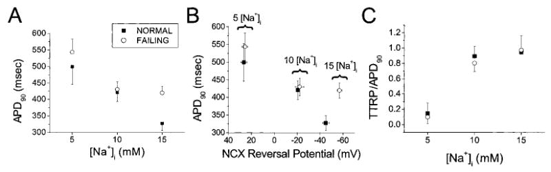
[Na+]i dependence of the APD, NCX driving-force RP, and TTRP in normal (solid square) and failing (open circle) myocytes. A and B, APD90 shortened with increasing [Na+]i (A), in correspondence with a shift in the NCX RP toward more hyperpolarized potentials (B). C, RP occurred later in the AP at higher [Na+]i. TTRP/APD90 is the TTRP normalized to APD90.