


Figure 7.
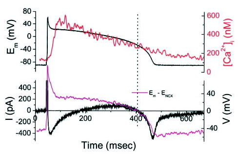
AP-clamp currents with 10 mmol/L [Na+]i and K+ currents blocked. Top panel shows AP waveform used to voltage-clamp a cardiomyocyte with Cs+ substitution of K+ in all solutions (to eliminate contamination by Ca2+ -activated Cl− current; this experiment was carried out in a guinea pig cardiomyocyte). Under these conditions, the predominant membrane currents were ICa,L and INCX. The kinetics and direction of the membrane currents during the AP plateau corresponded to estimates of the NCX driving force (magenta trace) calculated from the simultaneously recorded [Ca2+]i transient (red trace).